Anatomy and Clinical Significance of the Thoracic Cavity

A comprehensive overview of the thoracic cavity's organization, including its compartments (pleuropulmonary and mediastinum), thoracic wall, diaphragm, pleurae, and lungs. It also covers clinical correlations relevant to dentistry, such as referred pain, infection spread, and emergency management.

Save this reading to your study set

Sign up free

The thoracic cavity is a vital anatomical region that houses essential organs and provides structural protection. For healthcare professionals, particularly in dentistry, understanding this anatomy is critical for ensuring patient safety during examinations and treatments, as it helps in recognizing emergencies and understanding the spread of infections.

Organization of the Thoracic Cavity

The thoracic cavity is not a single empty space but is organized into three main functional compartments:

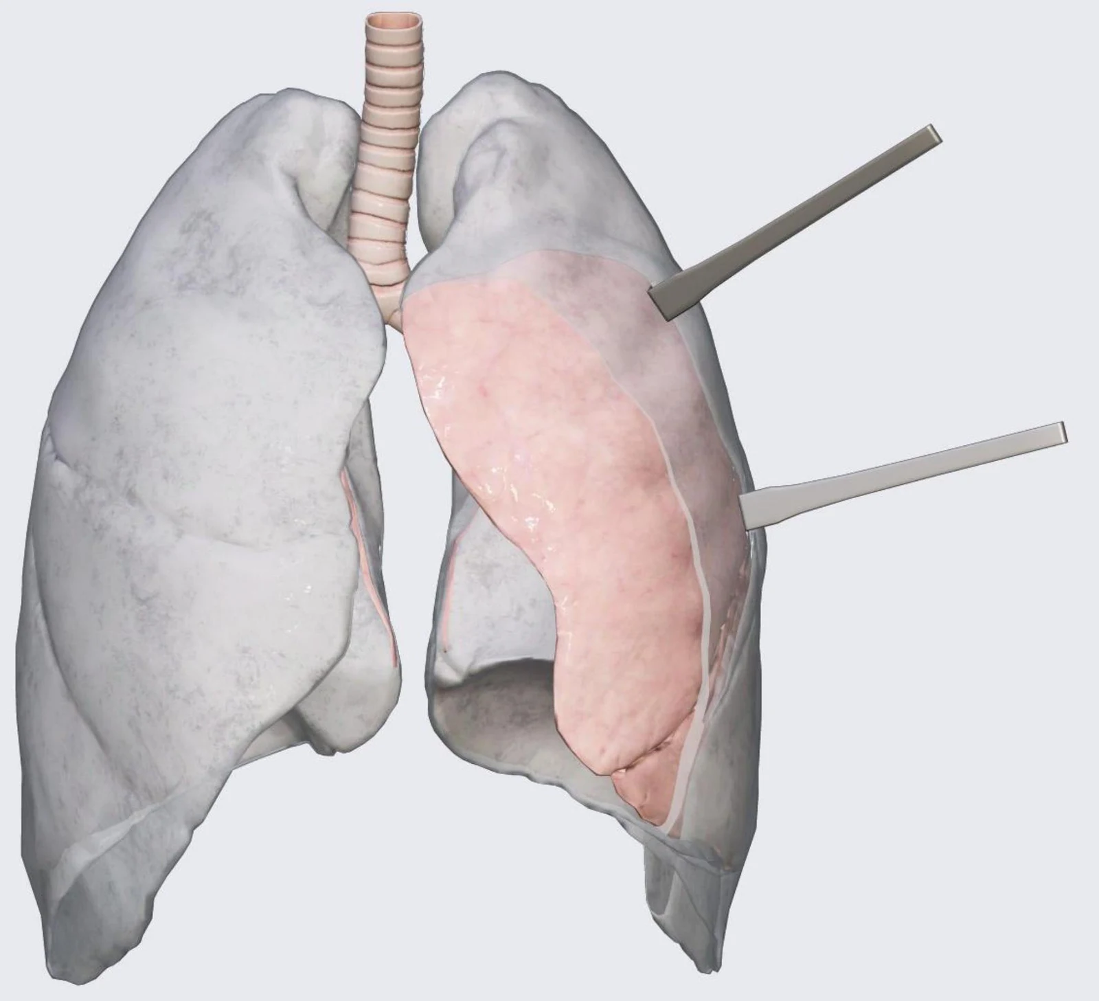
  1. Pleuropulmonary Compartments: Located on the right and left sides, these lateral compartments contain the lungs and their respective pleural sacs.
  2. Mediastinum: This is the central compartment situated between the two pleuropulmonary compartments. It contains the heart, pericardium, great vessels, trachea, oesophagus, lymphatics, and nerves.
  3. Superior Thoracic Aperture: This upper opening provides continuity between the thorax and the root of the neck.
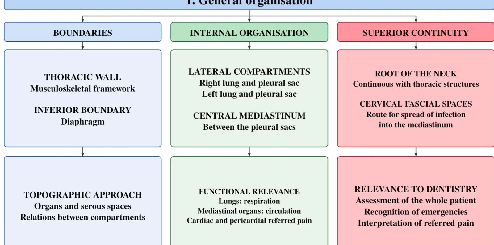
This diagram outlines the general organization of the thoracic region, detailing its boundaries, internal organization, and superior continuity. It further elaborates on the thoracic wall, inferior boundary, topographic approach, lateral compartments, central mediastinum, functional relevance, and relevance to dentistry.
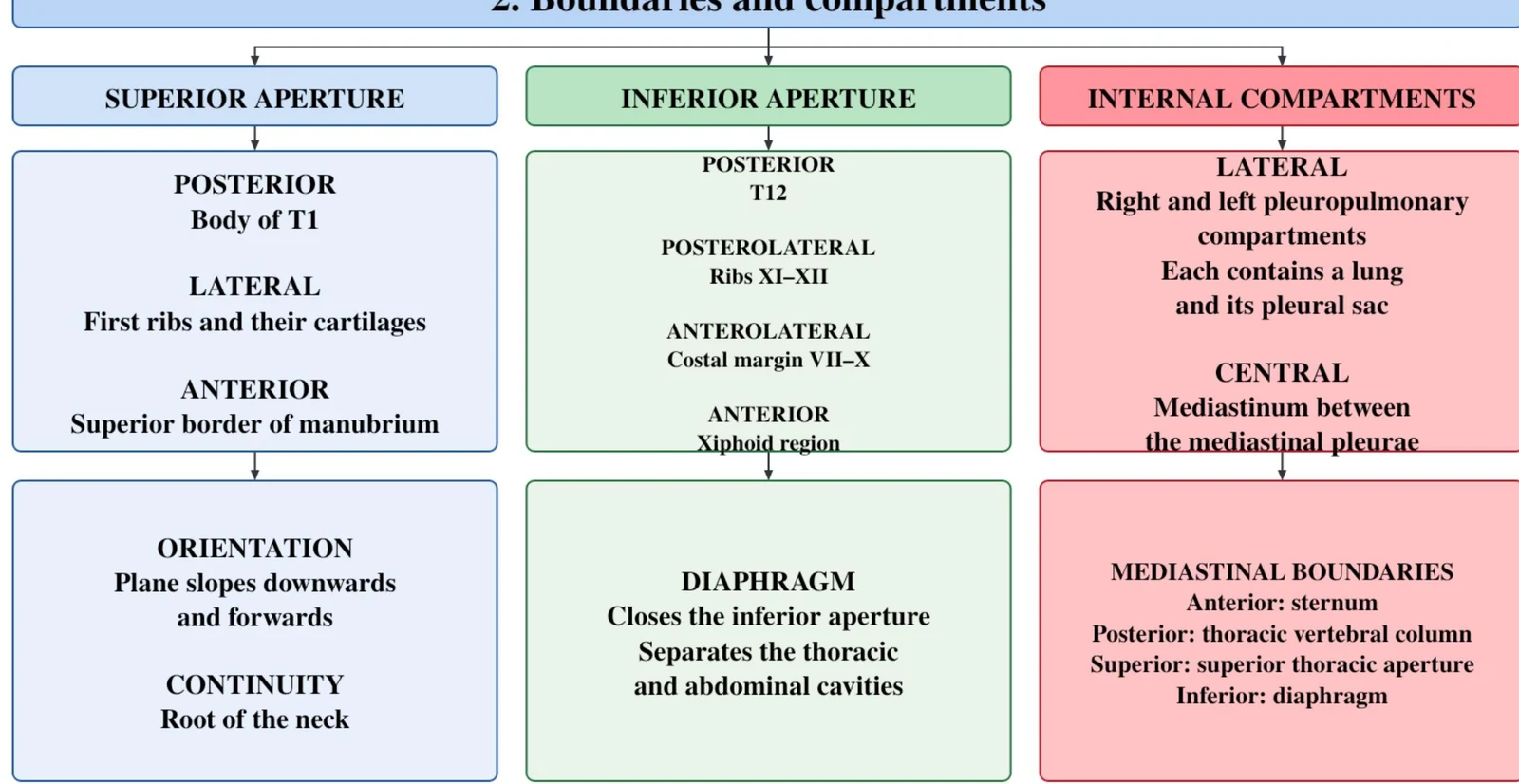
This diagram illustrates the boundaries and compartments of the thoracic region, including the superior and inferior apertures, internal compartments, diaphragm, and mediastinal boundaries.

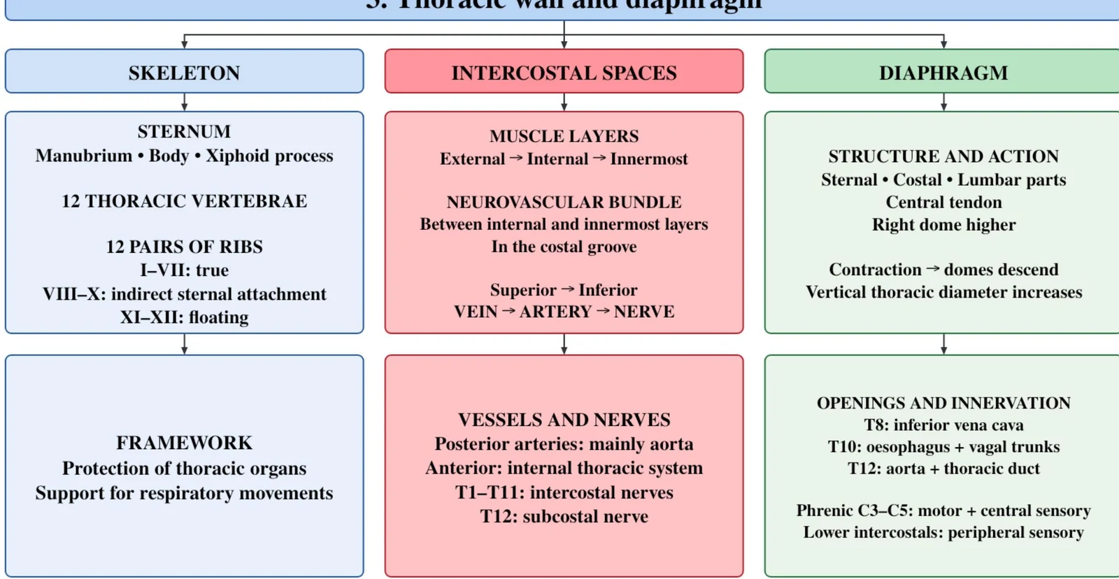
The Thoracic Wall and Diaphragm

The thoracic wall serves as a protective barrier for the internal organs and provides essential surface landmarks for clinical examination.

Surface Landmarks

A key landmark is the sternal angle. This structure is used to identify the second rib (rib II) and the transverse plane between the T4 and T5 vertebrae.
Keep reading for free.

Create an account to finish this reading and make it part of your study routine.

Continue reading free

Already have an account? Log in

Updated

Study it your way.

Import this reading into your study set to get started with another study mode.

Make it your own.

Keep this reading in a new study set, or add it to one you already use.