An in-depth exploration of the breast's anatomical structure, including its glandular tissue, supporting structures, and neurovasculature. It also covers lymphatic drainage pathways and clinical signs of malignant lesions.
The breast is a complex organ located within the subcutaneous tissue of the thoracic wall. While it is composed of various tissues including fat, lymphatics, and neurovasculature, its primary functional component is the mammary gland. Understanding the precise anatomical arrangement of these tissues, as well as the pathways for blood supply and lymphatic drainage, is essential for clinical practice, particularly in the diagnosis and management of malignant lesions.
Anatomical Structure and Composition
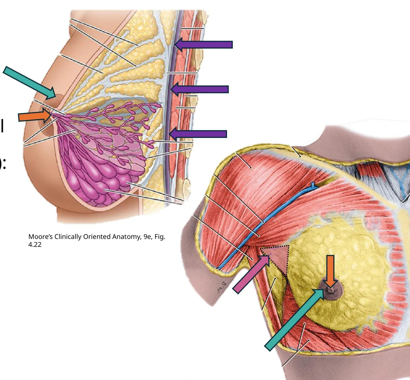
The breast lies upon the pectoral region, extending transversely from the lateral border of the sternum to the midaxillary line, and vertically from the 2nd to the 6th rib. The floor of the breast is formed primarily (two-thirds) by the pectoral fascia covering the pectoralis major muscle, with the remaining third formed by the fascia over the serratus anterior.
Histology and Glandular Tissue
The mammary gland is organized into 15–20 lobes. Within these lobes are smaller structures called lobules, which contain alveoli—the functional units that secrete milk. These lobes are drained by the lactiferous ducts, which lead toward the nipple. Just deep to the areola (the pigmented circular area of skin surrounding the nipple), the ducts widen into structures known as lactiferous sinuses.
Anatomical illustration of the female breast, showing glandular tissue, ducts, and surrounding musculature.
Supporting Structures
The breast is not merely a collection of glands; it is held in place by specialized connective tissue. The suspensory ligaments of Cooper are dense condensations of connective tissue that attach the lobes and lobules of the glands to the overlying skin. Additionally, the breast sits in a "retromammary space" located between the breast tissue and the pectoral fascia.
Clinical Note: Gynecomastia
Gynecomastia refers to the benign proliferation of glandular tissue in males. While temporary enlargement can occur during puberty due to hormonal shifts, persistent enlargement may indicate hormonal imbalances or metabolic changes in the liver.
Neurovasculature
The breast requires a robust supply of blood and nerve signals to maintain function and sensation.
Arterial Blood Supply
The blood supply to the breast is derived from several sources within the thoracic wall:
Medial mammary branches: Perforating branches from the internal thoracic artery.
Lateral branches: From the lateral thoracic and thoracoacromial arteries.
Posterior intercostal branches: Lateral mammary branches from the 2nd, 3rd, and 4th intercostal spaces.
Anatomical illustration of the thoracic aorta and its branches, including the posterior intercostal arteries and the internal thoracic artery with its perforating branches.
Venous Drainage
Venous blood is primarily drained via the lateral thoracic vein into the axillary vein. Some drainage occurs via the internal thoracic vein into the subclavian vein, while branches of the posterior intercostal veins drain into the azygos vein in the posterior thoracic wall.
Innervation
Sensory innervation to the breast and pectoral region is provided by the anterior and lateral cutaneous branches of the 4th through 6th intercostal nerves. Notably, the nipple serves as a key anatomical landmark for the T4 dermatome.
Lymphatic Drainage and Metastasis
Lymphatic pathways are of paramount clinical importance because they represent the primary route for the spread of breast cancer.
Primary Pathway: Lymph from the nipple, areola, and glandular tissue first passes to the subareolar lymphatic plexus. From there, more than 75% of the lymph drains to the axillary lymph nodes, then to the clavicular nodes, and eventually to the thoracic duct or right lymphatic duct.
Secondary Pathways: Less than 25% of lymph drains medially to the parasternal lymph nodes, to the opposite breast, or inferiorly to the subdiaphragmatic inferior phrenic lymph nodes.
Skin Drainage: Lymph from the skin (excluding the nipple/areola) drains into the ipsilateral axillary, infraclavicular, and deep cervical nodes.
Exam Tip: Metastatic Routes
Because of the interconnected nature of the lymphatic system, cancer can spread from the axillary nodes to the supraclavicular nodes, the opposite breast, or even the abdomen. Furthermore, because posterior intercostal veins drain into the azygos system, cancer can spread via the internal vertebral venous plexus to the vertebrae, cranium, and brain.
Clinical Pathology: Malignant Lesions
Types of Adenocarcinoma
Adenocarcinomas are malignancies that arise from the glandular tissues (the lobules or ducts). Two common types include:
DCIS (Ductal Carcinoma In Situ): Cancer located within the ducts.
LCIS (Lobular Carcinoma In Situ): Cancer located within the lobules.
Clinical Signs of Malignancy
Clinicians look for specific physical signs that suggest the presence of a tumor:
Peau d'orange: An "orange-peel" appearance of the skin. This occurs when cancer interferes with lymphatic drainage, causing lymphedema. The skin becomes puffy between dimpled pores and may feel thickened or leathery.
Skin Dimpling and Retraction: Large indentations (fingertip size or larger) can occur when a tumor invades the glandular tissue and causes fibrosis. This fibrosis can place traction on the suspensory ligaments of Cooper, pulling the skin inward.
Nipple Deviation: Malignant processes can cause the nipple to deviate from its normal position.
Invasion of Adjacent Tissue: If a tumor invades the retromammary space or the pectoral fascia, the breast may elevate when the pectoralis major muscle contracts.
Summary Table: Clinical Localization
Feature
Anatomical Cause
Peau d'orange
Lymphedema from blocked lymphatic drainage
Skin Dimpling
Fibrosis pulling on Suspensory Ligaments (Cooper)
Nipple Deviation
Tumor mass or tissue contraction
Breast Elevation
Invasion of Pectoralis Major or Pectoral Fascia
Keep reading for free.
Create an account to finish this reading and make it part of your study routine.