Anatomy and Clinical Correlations of the Thoracic Cavity
A comprehensive overview of the thoracic cavity, its subdivisions (pleuropulmonary compartments and mediastinum), thoracic wall, diaphragm, lungs, heart, great vessels, airway, esophagus, and nerves. It also details clinical correlations relevant to dental practice, such as aspiration, referred pain, and cardiorespirat
The thoracic cavity is the superior portion of the trunk, serving as the protective housing for the vital organs of respiration and circulation. Bounded by the osteomusculocutaneous thoracic cage and the muscular diaphragm, this space is essential for maintaining systemic homeostasis. For healthcare professionals, particularly those in dentistry, understanding the topography of the thorax is not merely an exercise in descriptive anatomy; it is a prerequisite for recognizing medical emergencies, understanding the spread of infection, and interpreting referred pain patterns.
Limits and Subdivisions of the Thoracic Cavity
The thorax is enclosed by the thoracic cage, which consists of the sternum, twelve pairs of ribs, and the thoracic vertebral column. The cavity communicates superiorly with the base of the neck through the superior thoracic aperture. This aperture is bounded posteriorly by the T1 vertebra, laterally by the first ribs, and anteriorly by the jugular notch of the manubrium.
Inferiorly, the cavity is separated from the abdominal cavity by the diaphragm, which closes the inferior thoracic aperture. The diaphragm is a musculotendinous septum that forms two domes; the right dome is typically higher than the left due to the presence of the liver.
Topographically, the thoracic cavity is divided into three main compartments:
Two lateral pleuropulmonary compartments: Each contains a lung enclosed within a pleural sac.
The mediastinum: The central compartment located between the two pleuropulmonary compartments.
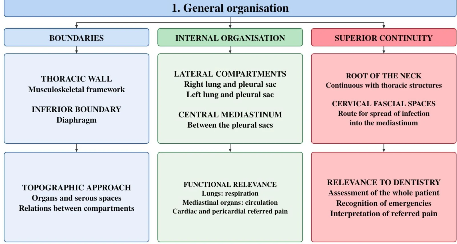
This diagram illustrates the general organization of the thoracic region, detailing its boundaries, internal organization, and superior continuity. It further elaborates on the thoracic wall, inferior boundary, lateral compartments, central mediastinum, topographic approach, functional relevance, and relevance to dentistry.
The Thoracic Wall and Diaphragm
The skeleton of the thorax provides both protection and the mechanical framework for respiration. The sternum is composed of the manubrium, the body, and the xiphoid process. A critical surface landmark is the sternal angle (or angle of Louis), which marks the articulation of the second costal cartilage. This landmark is used clinically to identify the second rib and the T4–T5 intervertebral plane.
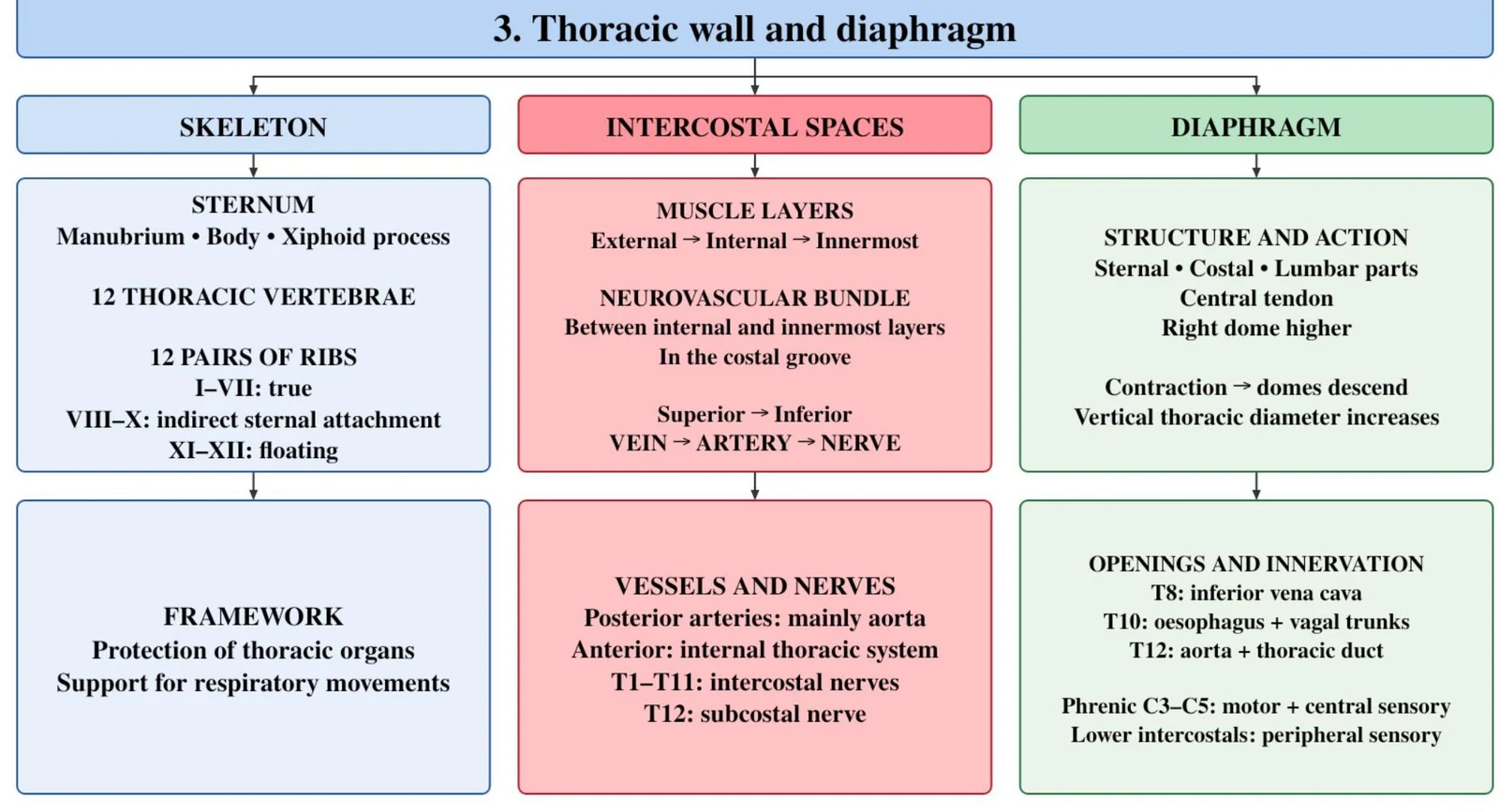
The intercostal spaces contain three layers of muscle (external, internal, and innermost), with the intercostal neurovascular bundle running in the costal groove at the inferior border of each rib. The order of structures from superior to inferior is vein, artery, and nerve.
The diaphragm is the primary muscle of inspiration. Its motor innervation is provided by the phrenic nerves ($C3$–$C5$), while the central portion receives sensory innervation from these nerves. The diaphragm features three major openings (hiatuses) that allow structures to pass between the thorax and abdomen:
Caval opening (T8): For the inferior vena cava.
Oesophageal hiatus (T10): For the oesophagus and vagal trunks.
Aortic hiatus (T12): For the aorta and thoracic duct.
This diagram outlines the components of the thoracic wall and diaphragm, including the skeleton, intercostal spaces, and diaphragm itself. It details structures like the sternum, ribs, muscle layers, neurovascular bundles, and their functions.
Pleurae and Lungs
The lungs are enclosed by a double-layered serous membrane called the pleura. The visceral pleura adheres directly to the lung surface, while the parietal pleura lines the thoracic wall, diaphragm, and mediastinum. The potential space between these layers is the pleural cavity, which contains a thin film of serous fluid.
The lungs are asymmetrical:
Right Lung: Shorter and wider (due to the high diaphragm); contains three lobes (superior, middle, inferior).
Left Lung: Contains two lobes (superior, inferior) and features a cardiac notch and a lingula.
Each lung is organized into bronchopulmonary segments, which serve as functional and surgical units. The lungs possess a double blood supply: the functional circulation (pulmonary arteries/veins) for gas exchange, and the nutritive circulation (bronchial arteries).
A box-and-connector summary detailing the anatomy of the pleurae, lungs, and their associated root and circulation. This is useful for respiratory system anatomy.
The Mediastinum and Its Contents
The mediastinum is a mobile connective tissue space bounded by the sternum anteriorly and the vertebral column posteriorly. It is divided by the horizontal plane of the sternal angle (T4–T5) into the superior and inferior mediastinum. The inferior mediastinum is further subdivided into anterior, middle, and posterior parts.
Subdivisions and Contents
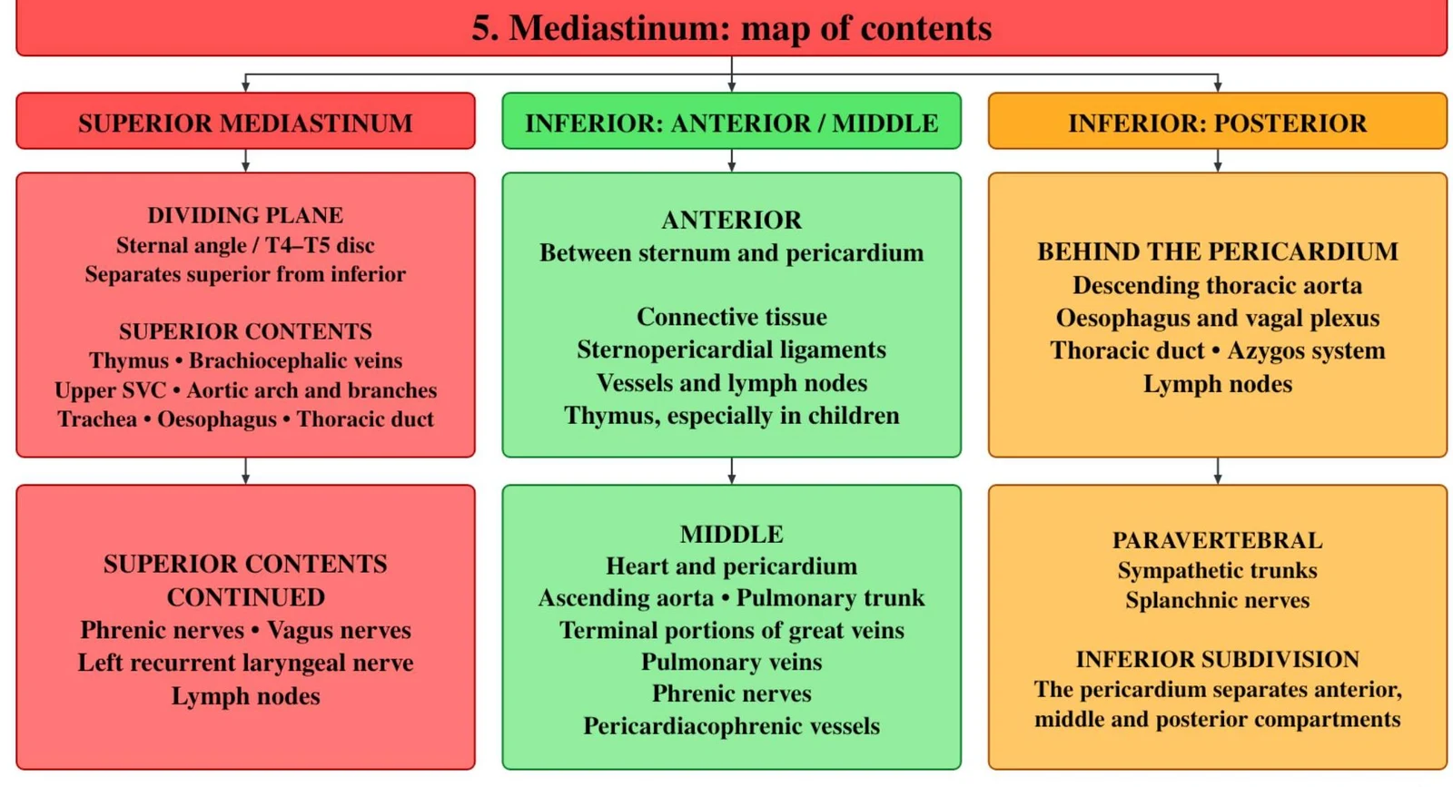
Superior Mediastinum: Contains the thymus, aortic arch, trachea, oesophagus, thoracic duct, and both phrenic and vagus nerves.
Anterior Mediastinum: A narrow space containing loose connective tissue and the inferior thymus in children.
Middle Mediastinum: Contains the pericardium, the heart, and the roots of the great vessels.
Posterior Mediastinum: Located between the pericardium and vertebrae; contains the descending aorta, oesophagus, vagus nerves, and thoracic duct.
Clinical Note on Infection Spread: The loose connective tissue of the mediastinum is continuous with the deep cervical fascial spaces of the neck. Consequently, odontogenic (dental) infections can descend into the thorax, potentially causing life-threatening descending mediastinitis.
This diagram illustrates the anatomical divisions and contents of the mediastinum, a region in the chest cavity.
Heart, Pericardium, and Great Vessels
The heart is situated in the middle mediastinum, enclosed by the pericardium. The pericardium consists of an inextensible fibrous layer and a serous layer (parietal and visceral/epicardium).
The heart functions as a pump, with blood flowing from the venae cavae to the right atrium, through the right ventricle to the pulmonary trunk, then to the lungs, back to the left atrium, and finally through the left ventricle into the aorta. The coronary arteries supply the myocardium and fill primarily during diastole.
The aortic arch is a major landmark, giving rise to three branches: the brachiocephalic trunk, the left common carotid artery, and the left subclavian artery.
A flowchart summarizing the anatomy, blood flow, and conduction system of the pericardium and heart. This is useful for cardiovascular physiology and medical anatomy.
Airway and Oesophagus
The trachea descends from the larynx and bifurcates into the main bronchi at the T4–T5 level. The carina, located at the bifurcation, is highly sensitive and triggers the cough reflex.
Aspiration Risk: The right main bronchus is shorter, wider, and more vertical than the left. Therefore, aspirated foreign bodies (such as dental crowns or instruments) are more likely to enter the right lung.
The oesophagus passes through the diaphragm at the T10 level. It features four physiological constrictions where swallowed foreign bodies may become lodged.
This diagram illustrates the anatomy and characteristics of the trachea, esophagus, and thymus, including their locations, relations, constrictions, supply, and age-related changes.
Nerves and Referred Pain
The thoracic cavity contains several critical nerve pathways:
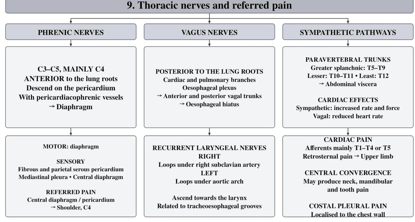
Phrenic Nerves ($C3$–$C5$): Pass anterior to the lung roots and provide motor supply to the diaphragm.
Vagus Nerves: Pass posterior to the lung roots and provide autonomic innervation to the heart, lungs, and oesophagus.
Referred Pain is a vital concept for clinical diagnosis. Because visceral afferent fibres from the heart enter the spinal cord at the same levels ($T1$–T4/T5) as somatic afferents from the neck, mandible, and teeth, cardiac pain can be perceived in the orofacial region.
Exam Tip: If a patient presents with diffuse, bilateral mandibular pain associated with exertion, sweating, or dyspnoea, consider a cardiac origin rather than a dental one.
This diagram illustrates the anatomy and function of thoracic nerves, including the phrenic nerves, vagus nerves, and sympathetic pathways, and their relation to referred pain.
Clinical Correlations for Dental Practice
For the dental professional, thoracic anatomy is directly linked to patient safety and emergency management:
Aspiration and Swallowing: Patients in semi-sitting positions are at risk of aspirating dental materials. Due to the anatomy of the right bronchus, these objects often lodge there.
Oral Bacteraemia: Bacteria from the oral cavity can enter the systemic circulation via the venous system, passing through the right heart and lungs to the left heart. In predisposed patients, this can lead to the colonization of cardiac valves (infective endocarditis).
Cardiorespiratory Emergencies: During emergencies like myocardial infarction, the patient's position is critical. Lying flat may worsen dyspnoea (orthopnoea) in patients with heart or lung disease. During CPR, compressions should be applied to the lower half of the sternum.
Local Anaesthesia: Adrenaline in local anaesthetics acts on cardiac β1 receptors, increasing heart rate and contraction force. Practitioners must assess cardiac risk and avoid intravascular injection.
Keep reading for free.
Create an account to finish this reading and make it part of your study routine.