


Permanent molars are the largest teeth in the human dentition, specifically designed for the heavy grinding required during mastication. Located distal to the premolars, these teeth play a critical role in maintaining functional occlusion, speech, and vertical facial dimension. Unlike premolars, permanent molars are nonsuccedaneous, meaning they do not replace any primary teeth but rather erupt into the space distal to the primary second molars.
Understanding the specific anatomical nuances of both maxillary and mandibular molars is not merely an exercise in memorization; it is a clinical necessity. Molar morphology directly dictates caries-risk assessment, the complexity of periodontal instrumentation, and the safety of surgical extractions.
General Molar Characteristics
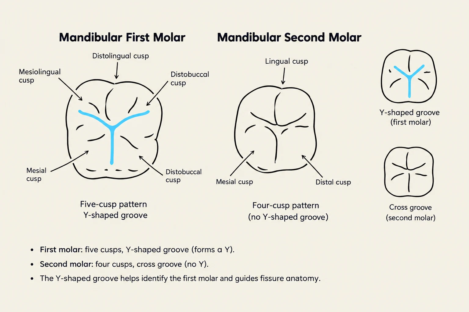
While molars vary by arch, they share certain fundamental properties. They possess broad occlusal surfaces characterized by multiple cusps, developmental grooves, supplemental grooves, fossae, and pits. The depth and complexity of these pits and grooves are primary factors in biofilm retention and subsequent caries risk.
NBDHE Exam Tip: Remember that permanent molars are nonsuccedaneous. They do not replace primary teeth; they erupt into the posterior space of the arch.
Maxillary Molars
Maxillary molars are generally wider buccolingually than they are mesiodistally. Most maxillary molars are trifurcated, possessing three roots: the mesiobuccal, distobuccal, and the lingual (or palatal) root. The lingual root is typically the largest and longest of the three.
The Maxillary First Molar
The maxillary first molar (#3 and #14) is the largest tooth in the maxillary arch and the largest crown in the entire permanent dentition. It is characterized by four major functional cusps, but a fifth minor cusp, known as the cusp of Carabelli, often appears on the lingual surface of the mesiolingual cusp. A defining anatomical feature is the oblique ridge, which crosses the occlusal table from the distobuccal cusp to the mesiolingual cusp.

Keep reading for free.
Create an account to finish this reading and make it part of your study routine.
Continue reading freeAlready have an account? Log in
Updated