Explores the distinct layers of the skin (epidermis, dermis, hypodermis), their cellular compositions, and their critical role in clinical practices such as injection techniques. It also covers accessory structures like hair, nails, and glands.
The skin, or integument, is the largest organ of the human body, accounting for approximately 16% of total body weight. While often discussed in terms of appearance, its primary physiological role is to act as a protective covering that separates the internal environment from the external world. Understanding the distinct layers of the skin is not merely an exercise in anatomy; it is a critical requirement for clinical practice, particularly when determining the correct technique for various types of injections.
The Layers of the Integument
The skin is composed of several distinct layers, each with unique cellular compositions and vascularity. These layers dictate how substances are absorbed and how the body responds to injury or medical intervention.
The Epidermis
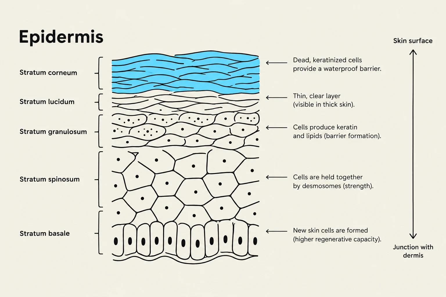
The epidermis is the most superficial layer of the skin. It is composed of stratified squamous epithelium, which acts like a "brick wall" of cells to provide a tough, resistant barrier. A key characteristic of the epidermis is that it is avascular, meaning it contains no blood vessels. Because of this, cells in the upper layers must rely on the diffusion of oxygen and nutrients from the underlying dermis.
The epidermis contains several specialized cell types:
Keratinocytes: These cells produce keratin, a waxy protein that provides water resistance and structural integrity.
Melanocytes: Located in the deepest layer (stratum basale), these cells produce melanin, the pigment responsible for skin color and UV protection.
Dendritic Cells: Phagocytic cells that act as part of the immune system by "eating" debris and pathogens.
Tactile Cells: Sensory cells associated with neurons that allow the skin to be highly sensitive to touch.
Diagram illustrating the layers of the epidermis, including the stratum corneum, stratum lucidum, stratum granulosum, stratum spinosum, and stratum basale, with descriptions of their functions.
The Dermis
Directly beneath the epidermis lies the dermis, a layer composed primarily of connective tissue. Unlike the epidermis, the dermis is highly vascularized, containing the blood vessels and nerves that support the skin. The dermis is divided into two layers: the thin, superficial papillary layer and the deeper, thicker reticular layer, which provides strength and elasticity through dense irregular connective tissue.
The Hypodermis
The layer below the dermis is the hypodermis, also known as the subcutaneous layer. This layer is composed largely of adipose tissue (fat). While not technically part of the skin itself, the hypodermis is vital because it anchors the skin to the underlying structures of the body and provides cushioning and impact resistance.
Clinical Injection Techniques
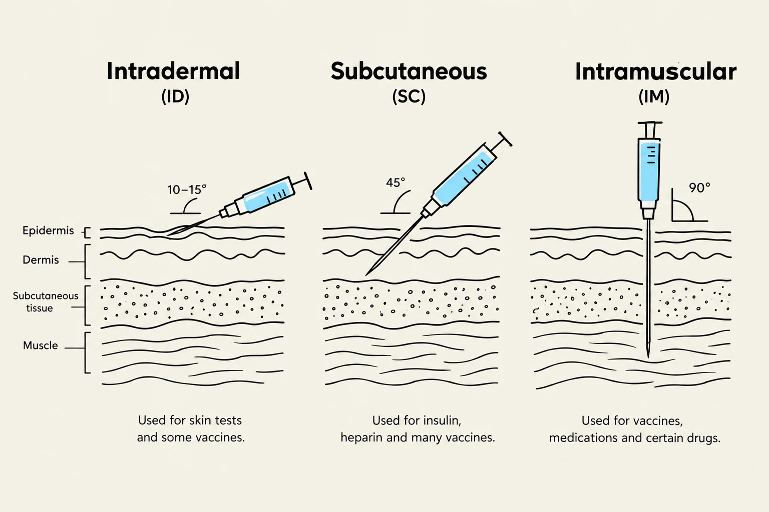
In a clinical setting, the depth and angle of a needle are determined by which layer of the skin is being targeted. The vascularity of these layers significantly affects how quickly a medication enters the systemic circulation.
Injection Type
Target Layer
Typical Angle
Clinical Use/Notes
Intradermal
Dermis
Shallow
Used for allergy testing to observe localized skin reactions.
Subcutaneous
Hypodermis (Adipose)
45∘
Targets the fat layer. Absorption is slower due to lower blood supply in adipose tissue.
Intramuscular
Skeletal Muscle
90∘
Penetrates epidermis, dermis, and hypodermis. Rapid absorption due to high vascularity of muscle.
Intravenous
Veins
Variable
Delivers substances directly into the bloodstream for the fastest administration.
Clinical Tip: When performing an intramuscular injection, common sites include the gluteus medius, deltoids, and lateral quadriceps. Always consider the patient's BMI, as a thicker layer of adipose tissue may require a longer needle to ensure the tip reaches the muscle.
Diagram illustrating intradermal, subcutaneous, and intramuscular injection techniques, including needle angles and typical uses.
Accessory Structures of the Skin
The skin includes several accessory structures that work in conjunction with the cutaneous membrane to maintain homeostasis.
Hair and Nails
Hair is composed of strands of dead keratinocytes containing hard keratin. It consists of the visible hair shaft and the hair bulb located within the hair follicle.
Texture: The shape of the follicle determines hair texture; round follicles produce straight hair, oval produce wavy hair, and flat produce curly hair.
Functions: Hair provides protection (from UV rays and debris), sensory reception (detecting environmental changes), and thermoregulation. Thermoregulation is aided by the arrector pili muscles, which contract to make hair stand up, trapping air to maintain warmth.
Nails are specialized epidermal projections that assist in grasping and manipulating objects.
Glands
The skin contains various glands that are essential for protection and temperature control:
Sebaceous Glands: These glands are typically associated with hair follicles and secrete sebum, a lipid-rich substance. Sebum serves as a lubricant, provides waterproofing, and is bactericidal, meaning it inhibits bacterial growth. Notably, sebaceous glands are absent in "thick skin" (the palms and soles) to prevent loss of grip.
Sudoriferous (Sweat) Glands: These include eccrine glands, which are vital for thermoregulation through evaporative cooling.
Summary Table: Key Functions of Skin Layers
Layer
Primary Component
Key Function
Epidermis
Keratinocytes
Protection, water resistance, UV absorption
Dermis
Connective Tissue
Strength, elasticity, nourishment (blood/nerves)
Hypodermis
Adipose Tissue
Insulation, cushioning, anchoring
Keep reading for free.
Create an account to finish this reading and make it part of your study routine.