An in-depth exploration of the shoulder complex, focusing on the glenohumeral joint, its surrounding muscles (deltoid, teres major, rotator cuff), and their roles in movement and stability. Includes biomechanics, kinematics, and clinical correlations like tendinitis and injuries.
The shoulder complex is one of the most mobile regions of the human body, a feat made possible by the unique relationship between the humerus and the scapula. Understanding the muscles that act on the glenohumeral (GH) joint requires a distinction between extrinsic muscles, which connect the axial skeleton to the limb, and intrinsic muscles, which act directly on the joint to facilitate movement and stability.
Biomechanics of Shoulder Movement
The movement of the shoulder is not limited to a single joint; it involves a sophisticated coordination of multiple articulations.
Glenohumeral Kinematics
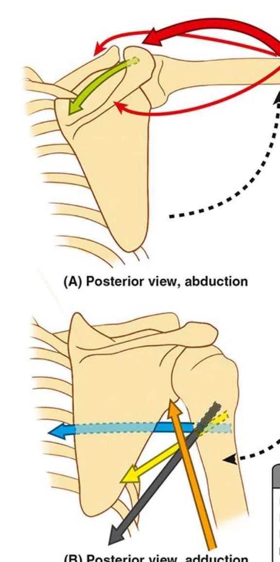
Glenohumeral kinematics refers to the movement occurring at the articulation between the head of the humerus and the glenoid cavity of the scapula. This includes flexion, extension, abduction, adduction, and rotation.
Diagrams illustrating shoulder abduction from a posterior view, showing the movement of the scapula and humerus.
Scapulothoracic Kinematics
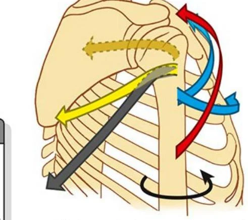
To achieve a full range of motion, the scapula must move against the rib cage. This is known as scapulothoracic kinematics. The coordination between the glenohumeral joint and the scapulothoracic joint is essential for efficient movement and preventing impingement.
Diagram illustrating the movements of the shoulder joint.
The Deltoid Muscle
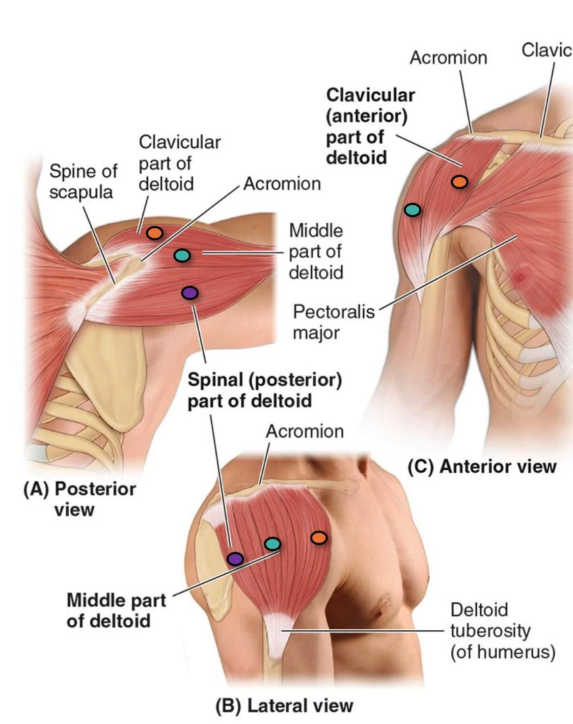
The deltoid is a large, powerful muscle that covers the shoulder joint. It is divided into three distinct parts, each contributing to different movements of the arm.
Feature
Description
Origin
Anterior: Lateral third of clavicle; Middle: Acromion of scapula; Posterior: Spine of scapula
When all three parts of the deltoid contract simultaneously, the primary action is abduction. It is important to note that the deltoid becomes the primary driver of abduction only after the first 15° of the movement.
Anatomical illustration of the human shoulder muscles, showing posterior, lateral, and anterior views with labels for the deltoid muscle parts, scapula, acromion, pectoralis major, and deltoid tuberosity.
The Teres Major
The teres major is an extrinsic muscle that assists in various movements of the humerus.
Origin: Posterior surface of the inferior angle of the scapula.
Insertion: Medial lip of the intertubercular sulcus of the humerus.
Action: Adduction and medial rotation; it also helps extend the arm from a flexed position.
Innervation: Lower subscapular nerve (C5, C6).
The Rotator Cuff (SITS Muscles)
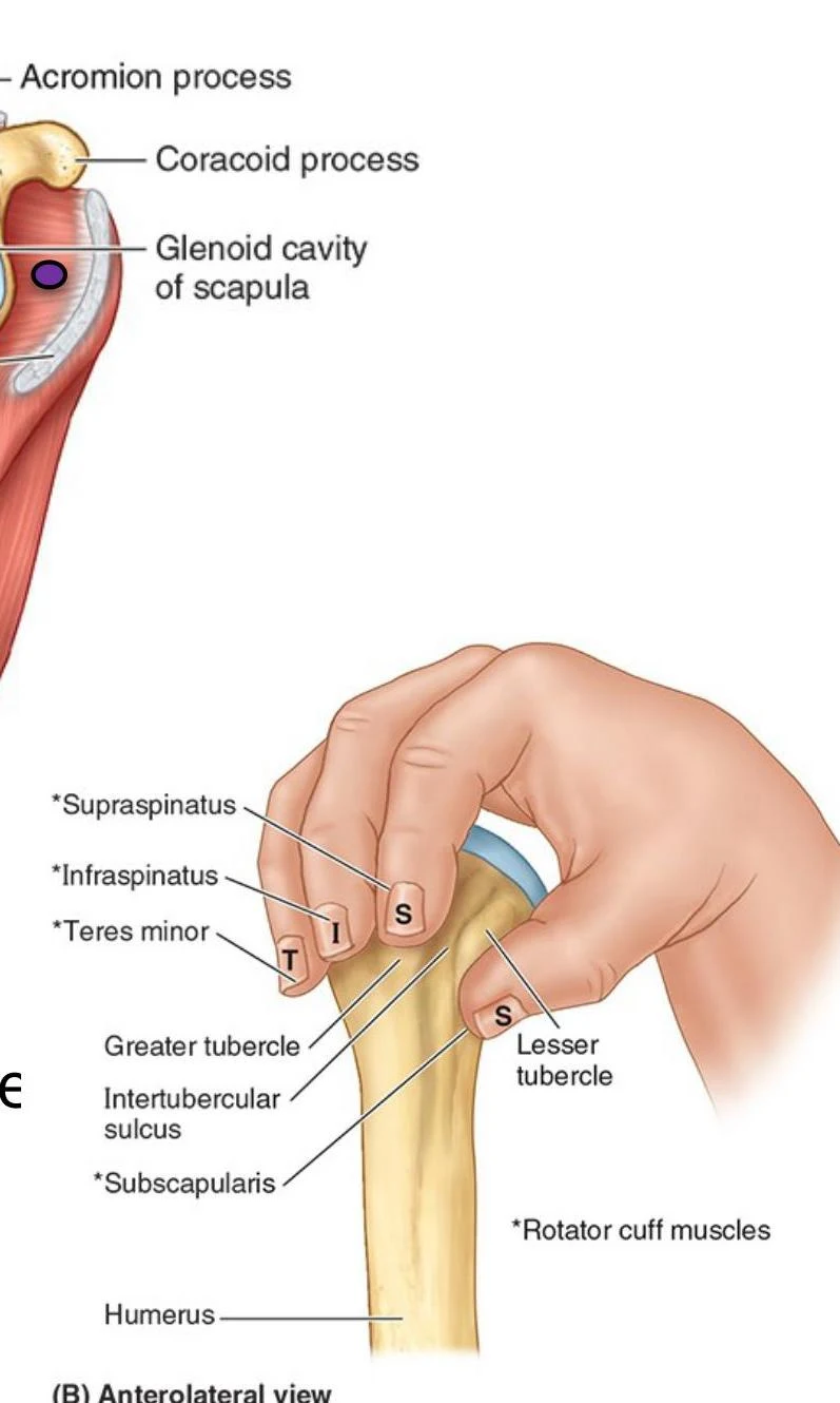
The rotator cuff is a functional unit of four muscles whose tendons fuse with the fibrous joint capsule. This creates a "musculotendinous cuff" that stabilizes the humeral head within the shallow glenoid cavity.
An illustration of the human shoulder joint, detailing the glenoid cavity of the scapula, humerus, and rotator cuff muscles including supraspinatus, infraspinatus, teres minor, and subscapularis.
The muscles are commonly remembered by the acronym SITS:
1. Supraspinatus
The supraspinatus originates in the supraspinous fossa of the scapula and inserts on the superior facet of the greater tubercle of the humerus. Its primary role is to initiate the first 15° of abduction and assist the deltoid thereafter. It also works with the other cuff muscles to keep the humeral head centered in the glenoid.
Innervation: Suprascapular nerve (C5, C6).
2. Infraspinatus
Located in the infraspinous fossa, this muscle inserts on the middle facet of the greater tubercle. Its primary action is lateral (external) rotation of the humerus.
Innervation: Suprascapular nerve (C5, C6).
3. Teres Minor
This muscle originates from the middle part of the lateral border of the scapula and inserts on the inferior facet of the greater tubercle. Like the infraspinatus, it performs lateral rotation and assists in adduction.
Innervation: Axillary nerve (C5, C6).
4. Subscapularis
Unlike the others, the subscapularis is located on the anterior aspect of the scapula (the subscapular fossa). It inserts on the lesser tubercle of the humerus and is the primary medial (internal) rotator of the arm.
Innervation: Upper and lower subscapular nerves (C5, C6).
Exam Tip: Rotator Actions
Remember that all rotator cuff muscles contribute to joint stability, but only the supraspinatus is not a rotator of the humerus. The others are categorized by whether they rotate the arm medially (subscapularis) or laterally (infraspinatus, teres minor).
Clinical Correlations
Supraspinatus Tendinitis and Impingement
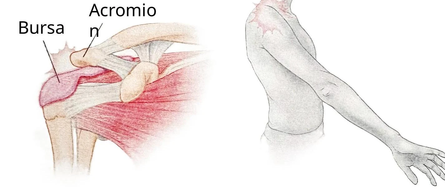
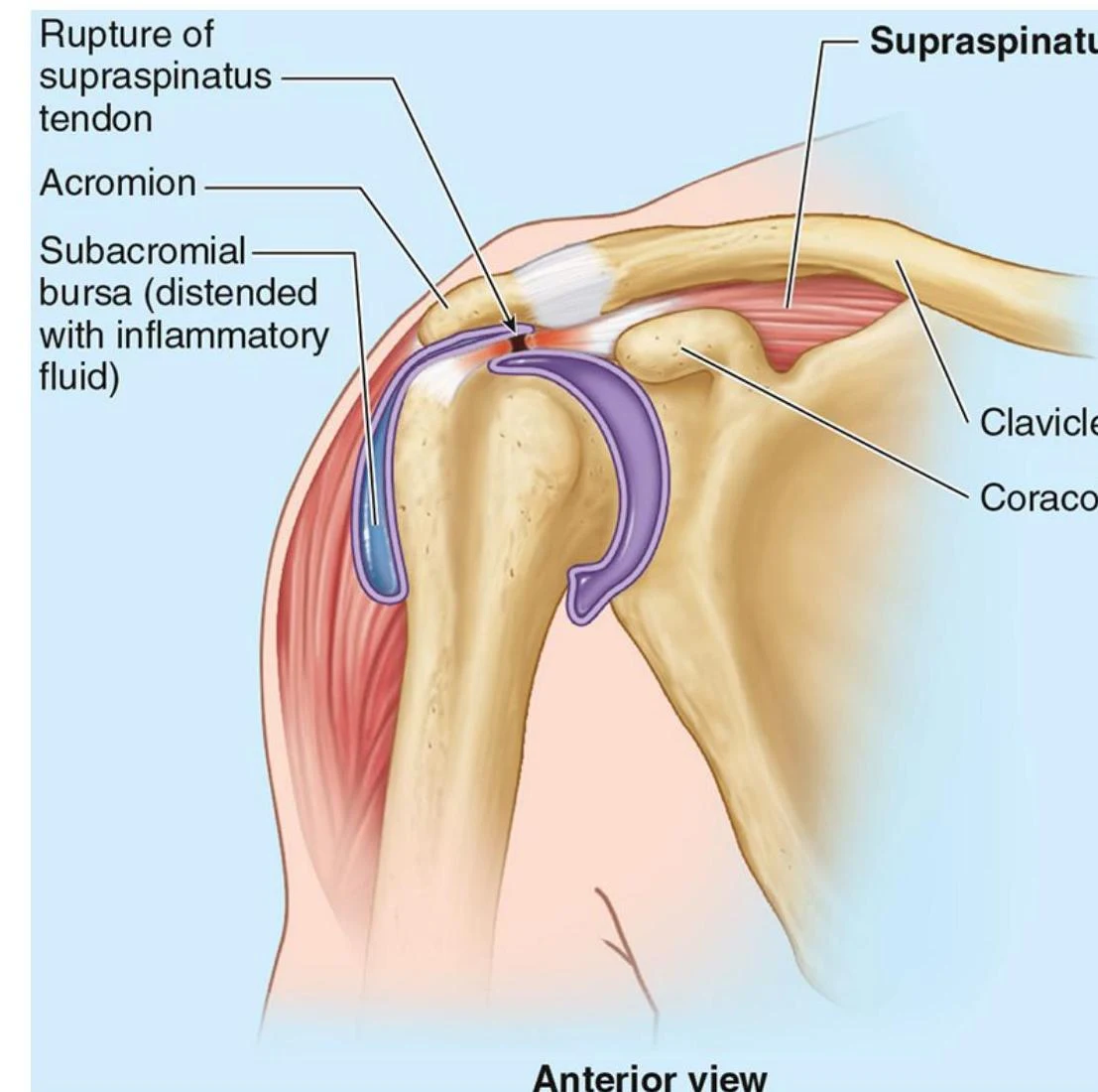
Because of its anatomical position, the supraspinatus tendon can become impinged between the acromion and the greater tubercle of the humerus. This often results from repetitive use or strain, leading to inflammation.
A key clinical diagnostic tool is the Painful Arc Test. A positive test occurs when a patient experiences pain during abduction between 60° and 120°, the specific range where the supraspinatus tendon is compressed against the inferior surface of the acromion.
An illustration of the human shoulder joint, highlighting the acromion and bursa.
Rotator Cuff Injuries
Rotator cuff injuries can stem from trauma, disease, or general wear and tear. The supraspinatus is the most commonly ruptured tendon. Patients typically present with:
Shoulder pain (often worsening at night).
Weakness when lifting or moving the arm.
Limited range of motion.
Joint crepitus (a grinding or popping sensation during movement).
An anterior view of the human shoulder illustrating a rupture of the supraspinatus tendon and a distended subacromial bursa with inflammatory fluid.
Quadrangular Space Syndrome
The quadrangular space is a passage between the teres minor (superiorly) and teres major (inferiorly). It contains the axillary nerve and the posterior circumflex humeral artery.
If the muscles forming this space become hypertrophied or fibrotic, they can impinge on the axillary nerve. This leads to "Quadrangular Space Syndrome," characterized by diffuse posterior shoulder pain, lateral arm paresthesia (numbness/tingling), and weakness in the deltoid.
Keep reading for free.
Create an account to finish this reading and make it part of your study routine.