


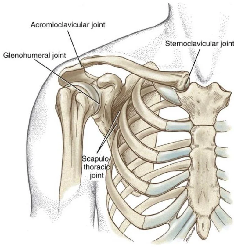
The shoulder complex is not a single joint, but a sophisticated functional unit designed to maximize mobility while maintaining stability. It integrates the clavicle, scapula, and humerus through several distinct joints, all of which are stabilized by an intricate web of ligaments, muscles, and neurovascular structures. Understanding this complex requires a multi-faceted approach, looking at the bones (osteology), the joints (arthrokinematics), the muscles (myology), and the neurological pathways (myotomes and dermatomes) that control them.
Neuroanatomy: Myotomes and Dermatomes
In clinical practice, assessing neurological integrity involves testing two distinct pathways: sensory and motor.
Dermatomes refer to the specific areas of skin supplied by afferent nerve fibers from the dorsal root of a single spinal nerve. For the upper extremity, key landmarks include:
- C4: The shoulder area.
- C5: The lateral arm extending to the proximal forearm.
- C6: The lateral forearm and the thumb.
- C7: Digits 2 and 3 (middle finger).
- C8: Digits 4 and 5 (ring and pinky fingers).
- T1: The medial forearm and elbow.
Myotomes represent the group of muscles innervated by a single spinal nerve root. If a nerve root is compressed, the corresponding muscle group may exhibit weakness. Key shoulder-related myotomes include:
- C4: Scapular elevation.
- C5: Shoulder abduction.
- C6: Elbow flexion, wrist extension, and forearm supination.
- C7: Elbow extension, wrist flexion, and forearm pronation.
- C8: Thumb extension and pronation.
Exam Tip: When testing for a C5 nerve root injury, focus on shoulder abduction strength. For a C6 injury, check elbow flexion and wrist extension.
Osteology and Joint Stability
The shoulder complex is composed of several bones and joints that work in concert to provide a wide range of motion.
Keep reading for free.
Create an account to finish this reading and make it part of your study routine.
Continue reading freeAlready have an account? Log in
Updated