


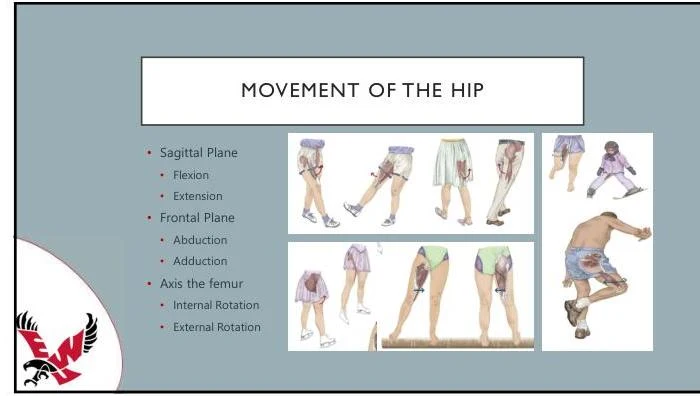
The pelvis and hip complex serves as the critical structural bridge between the axial skeleton (the spine) and the lower extremities. This region is responsible for weight-bearing, transferring forces during locomotion, and protecting vital pelvic organs. Understanding its anatomy requires a dual focus on the bony architecture of the pelvic girdle and the functional mechanics of the hip joint.
The Architecture of the Coxal Bones
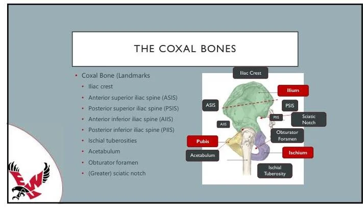
The primary structural unit of the pelvic girdle is the coxal bone (also referred to as the hip bone). While they may appear as single units in adults, the coxal bones are actually formed by the fusion of three distinct bones: the ilium, the ischium, and the pubis.
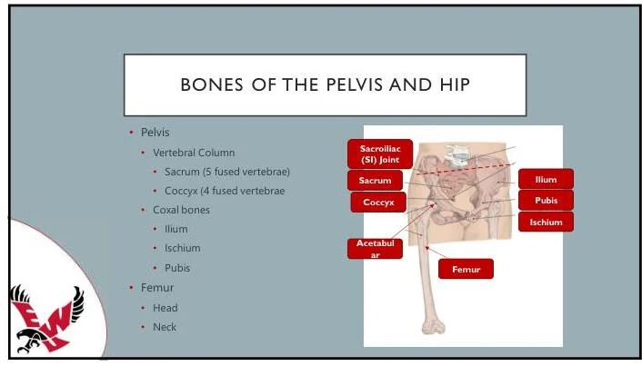
- Ilium: The largest and most superior component of the coxal bone, forming the broad, flared upper portion of the pelvis.
- Ischium: The posteroinferior portion of the bone, often referred to as the "sit bone" because it supports the body's weight when seated.
- Pubis: The anterior portion of the bone that meets its counterpart from the opposite side at the pubic symphysis.
Where these three bones meet, they form a deep, cup-shaped socket known as the acetabulum. The acetabulum acts as the "socket" in the ball-and-socket hip joint, receiving the head of the femur to allow for a wide range of motion.
Bony Landmarks and Pelvic Orientation
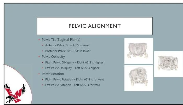
To assess the position and health of the pelvis, clinicians look to specific bony landmarks. These landmarks provide a roadmap for understanding how the pelvis is oriented in space.
Keep reading for free.
Create an account to finish this reading and make it part of your study routine.
Continue reading freeAlready have an account? Log in
Updated