


The human hand is a masterpiece of biomechanical engineering, allowing for both power grips and delicate fine motor tasks. This precision is made possible by a complex interplay of tendons, pulleys, and ligaments that stabilize the joints while facilitating smooth movement. Understanding the anatomy of the finger requires a deep dive into the structures that manage tension and the clinical consequences that arise when these systems fail.
Finger Ligaments and Pulleys
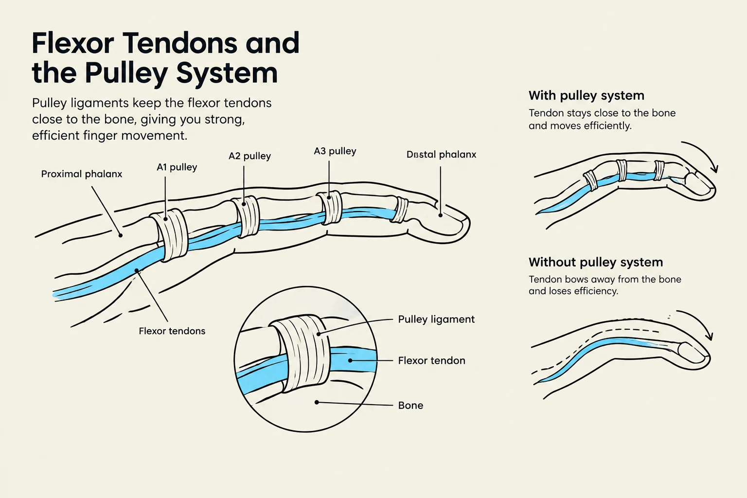
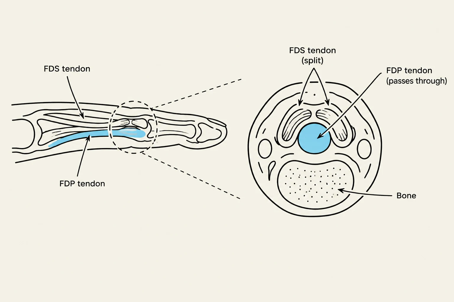
The stability of the finger during flexion is largely dependent on the relationship between the flexor tendons and the bone. Because the tendons are located on the palmar side of the phalanges, they have a natural tendency to "bowstring" away from the bone when tension is applied. To prevent this, the body utilizes a system of ligaments known as pulleys.
These pulleys act as anchor points that hold the flexor tendons close to the bone, ensuring that the force generated by the muscles is efficiently converted into joint rotation rather than wasted by pulling the tendon away from the shaft. Damage to these pulley systems can lead to significant loss of grip strength and mechanical efficiency.
The Extensor Hood and Cruciate Ligaments
While the palmar side of the finger is focused on flexion, the dorsal (top) side is specialized for extension and stability. The primary structure here is the extensor hood (or extensor expansion). This is a complex web of connective tissue that converges on the phalanges to allow for the coordinated extension of the Metacarpophalangeal (MP) and Interphalangeal (IP) joints.
Keep reading for free.
Create an account to finish this reading and make it part of your study routine.
Continue reading freeAlready have an account? Log in
Updated