An in-depth exploration of the cervical spine's unique articulations, ligaments, and musculature, detailing the structures responsible for head movement and stability.
The cervical spine is a marvel of biological engineering, designed to balance two competing needs: extreme mobility and structural stability. While the lower regions of the spine provide the heavy-duty support required for upright posture, the cervical region must allow for the wide range of motion necessary for vision and sensory orientation. This article explores the specialized joints of the upper neck, the complex network of ligaments that secure them, and the muscular layers that drive movement.
Intervertebral Articulations
The articulations of the cervical spine are categorized into two distinct groups: the specialized craniovertebral joints at the top of the column and the standard vertebral articulations found throughout the rest of the spine.
The Craniovertebral Joints
The uppermost part of the spine features two unique joints that facilitate the most significant movements of the head.
1. Atlantooccipital Joint
This is the first joint of the cervical spine, formed where the occipital condyles of the skull rest upon the articular fossa of C1 (the Atlas). It is classified as a double condyloid joint. Its primary function is to facilitate flexion and extension—often referred to as "capital flexion/extension" (the "yes" motion). While it allows for very slight lateral flexion and circumduction, its main role is the nodding motion of the head.
2. Atlantoaxial Joint
Located between C1 (Atlas) and C2 (Axis), this is a pivot joint. It is arguably the most important joint for head movement, as it is the most mobile joint between any two vertebrae. It facilitates the majority of transverse rotation (the "no" motion).
Diagrams illustrating the articulation of the occipital condyles with the atlas (C1) and axis (C2) vertebrae, showing front, side, and close-up views of the pivot joint.
General Vertebral Articulations
From the C2/C3 level down through the rest of the spinal column, the vertebrae articulate using two different types of joints:
Anterior joints (Amphiarthrodial): These involve the intervertebral disc, which acts as a shock absorber between the vertebral bodies.
Posterior joints (Gliding): These are formed by the facet joints, which allow for smooth, sliding movements between the articular processes.
Cervical and Spinal Ligaments
Ligaments act as the "straps" of the skeletal system, providing the tension necessary to prevent excessive or dangerous movement.
Upper Cervical Ligaments
The stability of the craniovertebral joints is maintained by a specific set of ligaments:
Atlanto-occipital Ligaments: These connect the occipital bone of the skull to the C1 vertebra.
Occipitoaxial Ligaments: These connect the occipital bone directly to the dens (the bony projection) of C2.
Atlantoaxial Ligaments: These connect the atlas to the axis.
Clinical Note: The Transverse Ligament
A critical component of the atlantoaxial complex is the Transverse Ligament (sometimes referred to as "Hangman's ligament"). This ligament connects the transverse processes of the atlas and serves the vital function of holding the dens of C2 in place. If this ligament is torn, the C1 vertebra can shift forward, potentially compressing the spinal cord and damaging vital nerves, which can be fatal.
Spinal Column Ligaments
Moving down the column, several longitudinal and transverse ligaments provide structural integrity:
Anterior Longitudinal Ligament: Runs the entire length of the spinal column along the anterior side; it is noted for being relatively weak.
Posterior Longitudinal Ligament: Runs the length of the column posteriorly and forms the anterior wall of the spinal canal.
Ligamentum Flavum: A strong ligament that connects successive laminae.
Interspinous Ligament: Runs between the spinous processes of adjacent vertebrae.
Supraspinous Ligament: Connects the dorsal tips of the spinous processes.
Ligamentum Nuchae: A specialized, thick supraspinous ligament that extends from the C7 vertebra to the occipital protuberance.
Diagrams illustrating the anatomy of the cervical spine, including side, partial, and axial views, highlighting ligaments, vertebral bodies, intervertebral discs, laminae, and the spinal cord.
Cervical Movements and Muscular Control
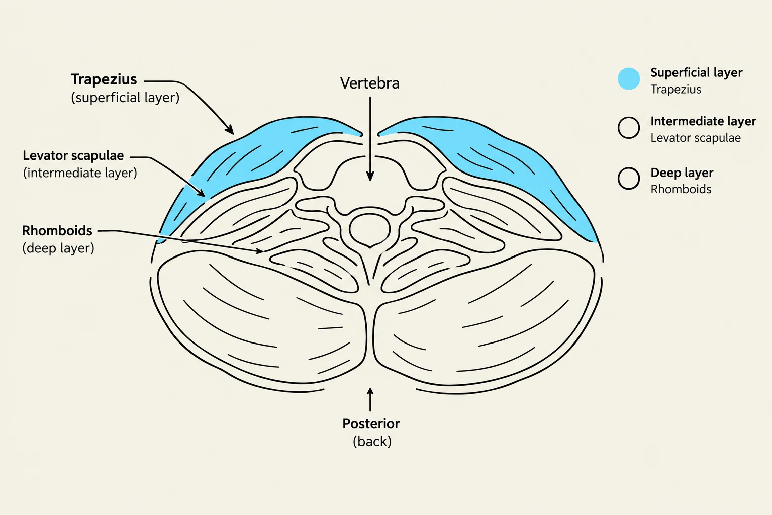
The movement of the cervical spine is a coordinated effort between the joints and the muscles organized into three distinct layers: superficial, intermediate, and deep.
Range of Motion
The typical ranges of motion for the cervical spine are:
Flexion: 45 degrees
Extension: 45 degrees
Rotation: 60 degrees
Lateral Flexion: 45 degrees
Muscular Layers of the Posterior Spine
1. Superficial Layer
This layer includes the Trapezius and Latissimus Dorsi, which contribute to general spinal movements. Deep to the trapezius are the Rhomboids (Major and Minor), which assist in spinal extension, lateral flexion, and rotation. Notably, the Rhomboid Minor and the Upper Trapezius are specifically involved in the rotation of the cervical spine.
2. Intermediate Layer: The Erector Spinae Group
The Erector Spinae (Sacrospinalis) is the largest muscle group, running parallel to the spinal column. They originate from the lumbar and sacral spinous processes and insert into various points including the ribs and transverse processes. This group is organized from lateral to medial using the mnemonic "I Like Standing":
Iliocostalis
Longissimus
Spinalis
Their primary actions are the extension, lateral flexion, and rotation of the spine.
3. Deep Layer
The deepest muscles provide fine-tuned control and stability:
Semispinalis Group (Capitis, Cervicis, Dorsi): These are responsible for the extension, lateral flexion, and rotation of the cervical spine.
Multifidus: These muscles facilitate both extension and rotation.
Quadratus Lumborum: Located in the lumbar region, this muscle performs extension and lateral flexion of the lumbar spine and assists in the depression of the 12th rib.
This diagram illustrates the superficial, intermediate, and deep layers of back muscles, including the Trapezius, Levator scapulae, and Rhomboids, in relation to a vertebra.
Study Tip: Movement Summary
When studying cervical movements, remember that rotation is primarily driven by the Atlantoaxial joint and supported by the Rhomboid Minor and Upper Trapezius, while flexion/extension (the "yes" motion) is the primary role of the Atlantooccipital joint.
Keep reading for free.
Create an account to finish this reading and make it part of your study routine.