


Mastering human anatomy requires more than just memorizing names; it demands an understanding of how these muscles interact to produce movement. For students preparing for practical exams, this involves identifying muscles on models, understanding their origins and insertions, and—most importantly—knowing their specific actions. This guide organizes essential muscle terms into functional groups, covering the shoulder, arms, legs, and the complex structures of the rotator cuff.
Understanding Muscle Tissue and the Sarcomere
Before diving into specific muscle groups, it is vital to understand the microscopic structures that allow for contraction. Muscle tissue is categorized into three types: skeletal, cardiac (heart), and smooth.
Skeletal muscle is characterized by the presence of endomysium, blood vessels, myofibers, motor neurons, motor end plates, nuclei, and sarcomeres. In contrast, cardiac muscle contains unique features like intercalated discs and autonomic neurons, while smooth muscle lacks the highly organized sarcomere structure seen in skeletal muscle.
At the functional unit level, the sarcomere is the fundamental unit of contraction. A detailed look at a giant sarcomere reveals several critical components:
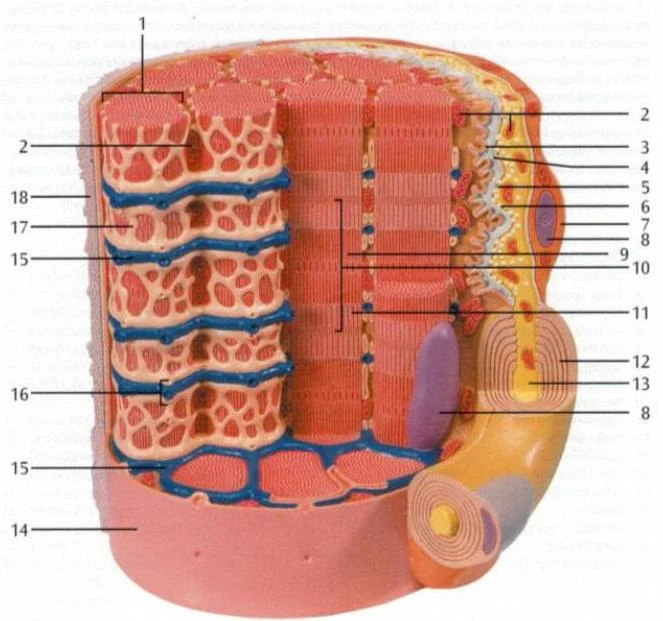
Key components of the sarcomere include:
- Z discs: The red ends that mark the boundaries of a single sarcomere.
- A band: The dark region containing both thick and thin filaments.
- I band: The light region containing only thin filaments.
- H zone: The central region of the A band containing only thick filaments.
- M line: The very center of the sarcomere.
- Filaments: The "thick" filaments are composed of myosin, while the "thin" filaments are composed of actin, supported by troponin and tropomyosin.
Keep reading for free.
Create an account to finish this reading and make it part of your study routine.
Continue reading freeAlready have an account? Log in
Updated