An Overview of the Integumentary, Lymphatic, and Peripheral Vascular Systems
This content provides a comprehensive overview of the human integumentary system (skin, hair, nails), the lymphatic system, and the peripheral vascular system, detailing their structures, functions, developmental considerations, and common clinical conditions.
The human body relies on a complex network of systems to maintain homeostasis, protect against environmental threats, and ensure the efficient transport of nutrients and waste. The integumentary system serves as our primary interface with the external world, providing a protective barrier through the skin, hair, and nails. Beneath this surface, the lymphatic system manages fluid balance and immune surveillance, while the peripheral vascular system facilitates the critical delivery of oxygenated blood and the return of deoxygenated blood to the heart. Understanding the anatomy, physiology, and developmental changes of these systems is essential for accurate clinical assessment.
The Integumentary System: Structure and Function
The integumentary system is much more than just a covering; it is a dynamic organ system responsible for protection, temperature regulation, perception, and the production of vitamin D. It minimizes injury from physical, chemical, and thermal sources while preventing the excessive loss of water and electrolytes.
Layers of the Skin
The skin is organized into three distinct layers, each with specialized roles:
Epidermis: This is the thin, tough, and avascular outer layer. It contains the basal cell layer, which is responsible for producing new keratinized cells and melanocytes (the cells that produce melanin), and the horny cell layer, which consists of dead, keratinized cells that are constantly shed and replaced approximately every four weeks.
Dermis: Located beneath the epidermis, this layer consists of connective tissue (collagen) and elastic tissue. It houses vital structures including nerves, sensory receptors, blood vessels, lymph vessels, and epidermal appendages.
Subcutaneous Layer: Also known as the hypodermis, this layer is composed of adipose (fat) tissue. It provides insulation, energy storage, cushioning, and allows for the mobility of the skin.
This diagram illustrates the three main layers of the skin: the epidermis, dermis, and hypodermis, along with their key components like blood vessels, collagen, elastin, and adipose tissue.
Skin Color and Pigmentation
Skin color is determined by three primary sources: melanin (brown), carotene (yellow-orange), and the underlying vascular bed (red-purple). While everyone possesses the same number of melanocytes, the amount of melanin produced varies based on genetics, hormones, and environmental factors. It is important to note that skin color can also be influenced by skin thickness and the presence of edema.
Epidermal Appendages
The skin contains several specialized appendages that contribute to its function:
Hair: These are threads of keratin that grow in cyclical active and resting phases. Vellus hair is fine and covers most of the body, whereas terminal hair is thicker and darker (e.g., scalp, eyebrows, and pubic hair).
Sebaceous Glands: These glands secrete sebum through hair follicles to lubricate the skin; they are found everywhere except the palms and soles.
Sweat Glands: There are two main types. Eccrine glands open directly onto the skin and produce a dilute saline sweat for thermoregulation. Apocrine glands are located in the axillae, anogenital area, and nipples; they become active at puberty and produce body odor when they interact with skin bacteria.
Nails: These are hard plates of keratin. The pink color of the nail comes from the underlying vascular nail bed, and the lunula (the white half-moon shape) overlies the nail matrix, which is protected by the cuticle.
Exam Tip: ABCDE Assessment
When assessing moles (nevi) for potential malignancy (melanoma), use the ABCDE criteria:
Asymmetry: Do the two halves match?
Border: Is it even and smooth, or irregular and notched?
Colour: Is it one even colour, or does it have multiple shades?
Diameter: Is it greater than 6 mm?
Evolution: Is it changing in size, shape, or colour?
Developmental Considerations
The integumentary and vascular systems undergo significant changes throughout the lifespan:
Adolescents: Increased activity in sebaceous and apocrine glands often leads to oily skin, acne, and increased body odor.
Pregnant Individuals: Hormonal changes can cause increased pigmentation (such as linea nigra on the abdomen or chloasma on the face), as well as vascular changes like striae gravidarum (stretch marks). Bilateral pitting edema and varicose veins are also common in the third trimester.
Older Adults: The skin undergoes "slow atrophy," characterized by a loss of collagen, elasticity, and subcutaneous fat. This leads to dry skin, a higher risk of shearing/tearing, and delayed wound healing. Furthermore, decreased sweat gland function increases the risk of heat stroke.
The Lymphatic and Peripheral Vascular Systems
While the integumentary system protects the exterior, the vascular and lymphatic systems manage the internal environment.
Peripheral Vascular System
The peripheral vasculature consists of arteries and veins:
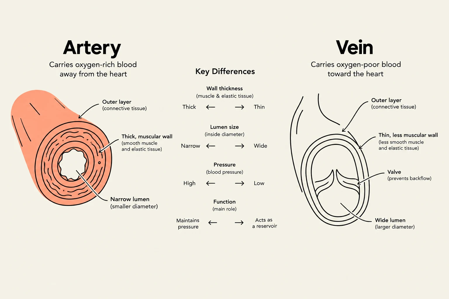
Arteries: These pump oxygenated blood to body tissues. Their walls contain elastic fibers to stretch during systole and muscle fibers to control blood flow diameter.
Veins: These return deoxygenated blood to the heart. Because they are a low-pressure system, they rely on skeletal muscle contraction, breathing pressure gradients, and intraluminal valves to ensure blood flow.
This diagram illustrates the key differences between arteries and veins, including their wall thickness, lumen size, pressure, and function in the circulatory system.
The Lymphatic System
The lymphatic system retrieves excess fluid from tissue spaces, filters it, and returns it to the bloodstream. It is divided into two main trunks: the Right Lymphatic Duct (draining the right side of the head, neck, chest, and right arm) and the Thoracic Duct (draining the rest of the body).
Key related organs include:
Spleen: Filters microorganisms and destroys old red blood cells.
Tonsils: Respond to local inflammation at the entrance of the respiratory and GI tracts.
Bone Marrow: The site of T and B lymphocyte creation.
Common Clinical Conditions
Edema: The accumulation of excess fluid in tissue. It is graded from 1+ (mild pitting) to 4+ (very deep pitting with significant swelling).
Peripheral Arterial Disease (PAD): Caused by atherosclerosis (fatty plaque buildup), leading to narrowed arteries and reduced blood flow.
Deep Vein Thrombosis (DVT): A blood clot in a deep vein, which can lead to a life-threatening pulmonary embolism.
Pressure Injuries: These occur when impaired circulation (often due to immobility) causes ischemia and cell death. They are staged from Stage 1 (intact skin with non-blanchable redness) to Stage 4 (full-thickness loss with exposed muscle or bone).
Clinical Note: Assessing Pulses
If a peripheral pulse is too weak to palpate manually, a Doppler Ultrasonic Stethoscope can be used. This device magnifies the pulsatile sounds from the blood vessels, allowing the clinician to locate the site via a "swishing" sound.
Keep reading for free.
Create an account to finish this reading and make it part of your study routine.