Alterations of Cardiovascular Function: A Comprehensive Overview
This content provides a comprehensive overview of cardiovascular diseases, covering cellular and tissue mechanisms, diseases of veins and arteries, vasculitis, hypertension, aneurysms, embolism, coronary artery disease, peripheral artery disease, and pediatric cardiovascular alterations.
The cardiovascular system is a complex network designed to maintain tissue perfusion and metabolic homeostasis. When this system is compromised by genetic, neurohumoral, or inflammatory mechanisms, the resulting alterations can lead to profound clinical consequences. Understanding these alterations requires a deep dive into the cellular processes of the endothelium, the structural changes in vessels, and the systemic responses to ischemia and pressure changes.
Diagram illustrating the stages of atherosclerotic plaque formation within an arterial wall, showing a healthy vessel, LDL penetration into the intima, and the development of an atherosclerotic plaque with a narrowed lumen.
Cellular and Tissue Mechanisms
At the heart of many cardiovascular diseases is endothelial injury. The endothelium is not merely a lining but a dynamic layer that regulates vascular tone and inflammation. Damage to this layer—caused by hypertension, smoking, hyperlipidemia, or toxins—triggers a cascade of events.
When the endothelium is injured, several key processes occur:
Atherosclerosis: This is a form of arteriosclerosis characterized by the accumulation of lipid-laden macrophages in the arterial wall. The process begins when LDL enters the intima and becomes oxidized, prompting monocytes to migrate and transform into "foam cells".
Remodeling: This refers to structural changes in the heart or vessels. In the context of hypertension, it involves hypertrophy and hyperplasia of the vessel walls. In the myocardium, remodeling occurs after a myocardial infarction (MI) as the heart attempts to repair itself through fibroblast proliferation and scar tissue synthesis.
Reperfusion Injury: This occurs when blood flow is restored to tissue after a period of ischemia, which can paradoxically cause further cellular damage.
Stunning: A temporary loss of contractile function that persists for hours or days even after perfusion has been restored.
Exam Tip: Distinguish between stunning (temporary loss of function) and hibernating myocardium (persistent ischemia where the tissue undergoes metabolic adaptation to survive).
Diseases of the Veins
Venous disorders primarily involve structural changes and obstructions to blood flow.
Varicose Veins and Chronic Venous Insufficiency
Varicose veins are distended, tortuous, and palpable veins, most commonly occurring in the saphenous vein. They are caused by trauma or gradual distention that renders the venous valves incompetent. This incompetence is often linked to altered connective tissue proteins and increased proteolytic enzyme activity. If the inadequate venous return persists, it leads to chronic venous insufficiency, which can manifest as venous stasis ulcers.
Superior Vena Cava Syndrome (SVCS)
SVCS is an oncologic emergency caused by the progressive occlusion of the Superior Vena Cava (SVC), most frequently by bronchogenic cancer. Patients present with:
Edema and venous distention of the face and upper extremities.
A "fullness" feeling in the head, headaches, and visual disturbances.
Skin on the face and arms appearing purple and taut.
Vasculitis
Vasculitis is the inflammation of blood vessels. It can be triggered by viral infections (such as Hepatitis B/C, EBV, or CMV), specific drugs (like Penicillin or Sulfa), or autoimmune diseases such as Systemic Lupus Erythematosus (SLE) and Rheumatoid Arthritis (RA).
Clinical manifestations include constitutional symptoms like fever, fatigue, and weight loss. If ischemia occurs, patients may suffer from skin ulcers, renal dysfunction, neuropathy, or bowel infarction. A definitive diagnosis typically requires an arterial biopsy or an angiogram.
Diseases of the Arteries
Arterial pathologies affect blood pressure, vessel patency, and tissue perfusion.
Hypertension
Hypertension is the consistent elevation of systemic arterial blood pressure.
Primary (Essential) Hypertension: Has no known cause and accounts for 95% of cases. It results from complex interactions between genetics and the environment, mediated by the sympathetic nervous system (SNS) and the renin-angiotensin-aldosterone system (RAAS).
Secondary Hypertension: Caused by an underlying disease, such as renal artery stenosis or drugs.
Orthostatic (Postural) Hypotension: A decrease in systolic BP by ≥20 mm Hg or diastolic BP by ≥10 mm Hg upon standing.
Aneurysms and Embolism
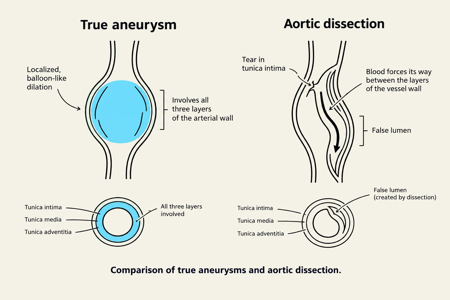
Comparison of true aneurysms and aortic dissection.
An aneurysm is a local dilation or outpouching of a vessel wall.
True Aneurysms involve all three layers of the arterial wall.
False Aneurysms occur when there is a leak between a vascular graft and a natural artery.
A critical complication is aortic dissection, a surgical emergency where a tear in the intima allows blood to flow into the vessel wall, furthering the tear.
An embolism is a bolus of matter (such as a dislodged thrombus, air bubble, or fat) that circulates in the bloodstream and lodges, obstructing flow. If an embolus occludes a coronary artery, it results in a myocardial infarction (MI); if it occludes a cerebral artery, it causes a stroke.
Coronary Artery Disease (CAD) and Myocardial Ischemia
CAD involves any vascular disorder that narrows or occludes the coronary arteries, usually due to atherosclerosis. This creates an imbalance between myocardial oxygen supply and demand.
Myocardial Ischemia: Occurs when the coronary blood supply cannot meet the metabolic demand. This can manifest as stable angina (predictable), Prinzmetal angina (unpredictable), or silent ischemia.
Acute Coronary Syndromes (ACS): Sudden coronary obstruction due to thrombosis over a ruptured plaque. This includes unstable angina (a harbinger of impending infarction) and Myocardial Infarction (MI).
In an MI, prolonged ischemia leads to irreversible myocyte necrosis. Clinical signs include sudden severe chest pain, diaphoresis, and dyspnea. Laboratory markers like Troponin I are highly specific and elevate within 2 to 4 hours of injury.
Peripheral Artery Disease (PAD)
PAD involves atherosclerosis of the arteries that perfuse the limbs, particularly the lower extremities. A common symptom is intermittent claudication (pain with ambulation). Severe cases may present with the "6 Ps": Pallor, pulselessness, pain, paresthesias, paralysis, and poikilothermia.
Pediatric Cardiovascular Alterations
Diagram illustrating parallel circulation in transposition of the great arteries, showing the flow of deoxygenated and oxygenated blood through the heart, lungs, and body.
In children, cardiovascular alterations are often linked to congenital heart defects. These are classified by their effect on blood flow:
Acyanotic Defects (Increasing Pulmonary Flow): Such as Patent Ductus Arteriosus (PDA), Atrial Septal Defect (ASD), and Ventricular Septal Defect (VSD). These often lead to pulmonary congestion and heart failure.
Cyanotic Defects (Decreasing Pulmonary Flow): Such as Tetralogy of Fallot, which involves four defects: VSD, overriding aorta, pulmonary valve stenosis, and right ventricular hypertrophy. These manifest with hypoxemia and cyanosis.
Obstructive Defects: Such as Coarctation of the aorta, a narrowing of the aortic lumen. This can cause hypertension in the upper extremities and decreased/absent pulses in the lower extremities.
Mixed Defects: Such as Transposition of the great arteries, where the aorta and pulmonary artery are switched, creating two separate, parallel circuits.
Clinical Note: In Kawasaki disease (a systemic vasculitis in children), the child is at highest risk for coronary artery aneurysm development during the subacute phase.
Keep reading for free.
Create an account to finish this reading and make it part of your study routine.