Alterations in Cardiovascular Function and Perfusion
This module provides an overview of the cardiovascular system, including heart structure, cardiac electrophysiology, hemodynamics, and assessment of heart sounds.
The cardiovascular system is a complex, integrated network designed to maintain homeostasis by delivering oxygen and nutrients to tissues while simultaneously removing metabolic waste products. This system relies on the continuous, coordinated action of the heart and the vast network of blood vessels, collectively known as the vascular system. When the heart's ability to pump or the vessels' ability to transport blood is compromised, it leads to alterations in cardiovascular function and perfusion, which can significantly impact a client's overall health.
Heart Structure and Layers
The heart is a hollow, muscular organ located in the mediastinal area of the chest, positioned between the sternum and the spinal cord. To function effectively, the heart is organized into distinct layers and chambers.
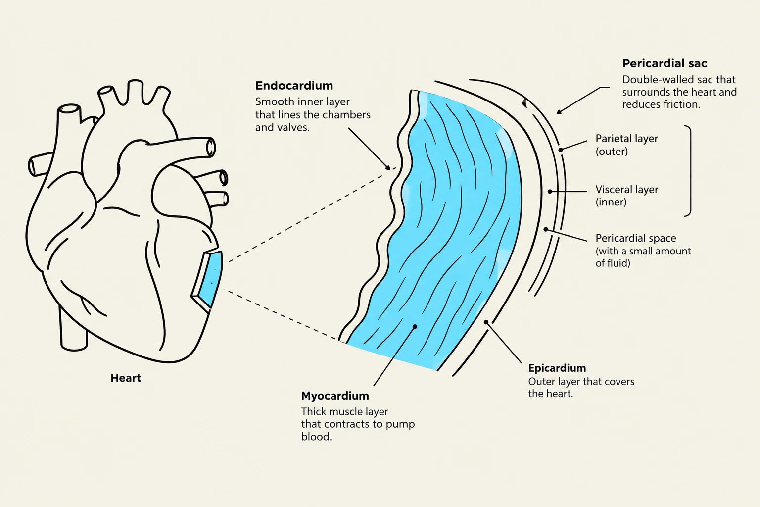
The heart wall consists of three primary layers:
Endocardium: The innermost layer made of endothelial tissue that lines the heart chambers and valves.
Myocardium: The thickest, middle layer composed of muscle fibers; it is the layer responsible for the heart's pumping action.
Epicardium: The outer serous layer that protects the heart.
Surrounding the entire organ is the pericardium, a fibrous sac. The pericardium includes the parietal pericardium (which lines the sac and anchors the heart) and the visceral pericardium (which covers the heart surface). Between these two layers lies the pericardial space, containing serous fluid that allows the layers to move smoothly against each other during contraction.
This diagram illustrates the layers of the heart, including the endocardium, myocardium, and epicardium, as well as the pericardial sac which surrounds the heart.
Heart Chambers and Valves
The heart is divided into four chambers: the right atrium, right ventricle, left atrium, and left ventricle. A band of tissue called the septum separates the right and left sides of the heart.
Blood flow follows a specific, unidirectional path regulated by four valves:
Atrioventricular (AV) valves: The tricuspid (right side) and mitral/bicuspid (left side) valves separate the atria from the ventricles.
Semilunar valves: The pulmonic and aortic valves separate the ventricles from the great arteries.
During diastole (relaxation), blood flows from the atria into the ventricles. During systole (contraction), the AV valves close to prevent backflow, and blood is ejected through the semilunar valves into the pulmonary artery or the aorta.
The Vascular System and Cardiac Perfusion
The vascular system is composed of three main types of vessels: arteries, veins, and capillaries.
Arteries: High-pressure vessels that transport blood away from the heart.
Veins: Low-pressure vessels that act as a reservoir and return blood to the heart.
Capillaries: Tiny vessels that connect arterioles to venules, serving as the site for gas, nutrient, and waste exchange between blood and tissues.
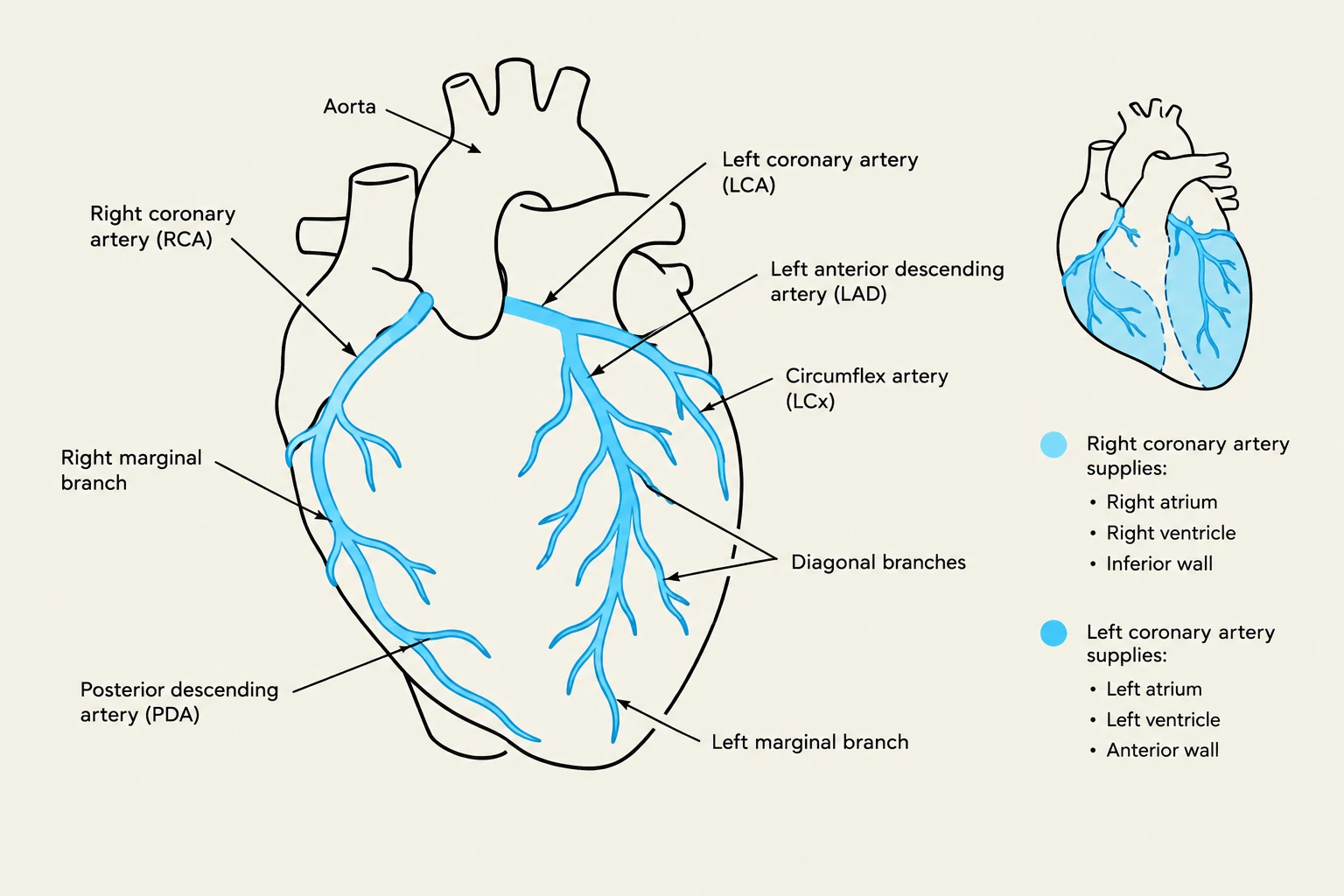
A critical component of cardiac perfusion is the coronary arteries, which originate at the base of the aorta and supply oxygen-rich blood directly to the myocardium. Notably, coronary perfusion occurs primarily during diastole; as the heart rate increases, the diastolic period shortens, which can decrease the time available for the heart muscle to receive its own blood supply.
Diagram illustrating the coronary arteries of the heart and their supply areas.
Cardiac Electrophysiology
The heart possesses an intrinsic conduction system that generates and transmits electrical impulses to trigger myocardial contraction. The sequence of an electrical impulse is as follows:
Sinoatrial (SA) Node: Located in the right atrium, it acts as the natural pacemaker, setting the heart's rate and rhythm.
Atrioventricular (AV) Node: Receives the impulse from the SA node; a slight delay here allows the atria to finish contracting and empty blood into the ventricles.
Bundle of His: Conducts the impulse from the AV node into the interventricular septum.
Purkinje Fibers: Located within the ventricular walls, these fibers distribute the impulse to ensure coordinated ventricular contraction.
An Electrocardiogram (ECG) is used to record this electrical activity. Key components include the P wave (atrial depolarization), the QRS complex (ventricular depolarization), and the T wave (ventricular repolarization).
Cardiac Hemodynamics and Output
Hemodynamics refers to the forces involved in circulating blood. The primary goal of the heart is to maintain an adequate cardiac output (CO), which is the total amount of blood ejected from a ventricle per minute.
The relationship between heart rate (HR) and stroke volume (SV) is expressed as:
Cardiac Output (CO)=Stroke Volume (SV)×Heart Rate (HR)
Stroke volume is determined by three critical factors:
Preload: The degree of stretching of the ventricular muscle at the end of diastole.
Afterload: The resistance or pressure the ventricle must overcome to eject blood into the circulation.
Contractility: The innate force of the myocardial contraction.
Exam Tip: If a client has hypertension, their afterload is increased because the resistance in the arterial system is higher, making it harder for the heart to pump blood out.
Clinical Assessment: Heart Sounds
Auscultation is a vital skill for detecting alterations in cardiac function. Normal heart sounds are produced by the closure of the valves:
S1 ("lub"): Closure of the mitral and tricuspid valves.
S2 ("dub"): Closure of the aortic and pulmonic valves.
Abnormal sounds include:
Murmurs: "Whooshing" sounds caused by turbulent blood flow through diseased or narrow valves.
S3 and S4: Extra sounds (gallops) that may indicate decreased ventricular compliance or heart failure.
Pericardial Friction Rub: A "grating" or "squeaky" sound caused by inflammation of the pericardial layers.
Summary of Dysrhythmias
When the conduction pathway is disrupted, dysrhythmias occur. Common examples include:
Sinus Bradycardia: A heart rate <60 bpm.
Sinus Tachycardia: A heart rate >100 bpm.
Atrial Fibrillation (A-fib): Characterized by irregular ventricular contractions, which can lead to an apical-radial pulse deficit.
Premature Ventricular Contractions (PVCs): Early contractions originating in the ventricles, often caused by electrolyte imbalances like hypokalemia or hypomagnesemia.
Keep reading for free.
Create an account to finish this reading and make it part of your study routine.