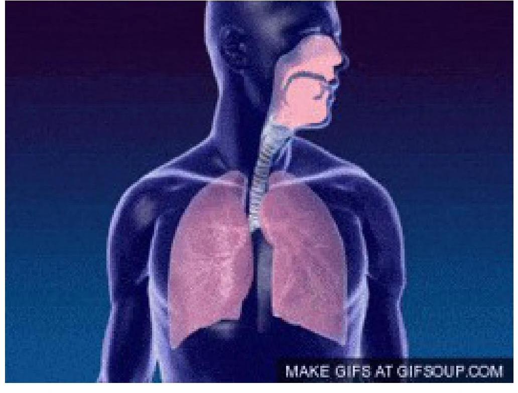
The alveoli are the tiny air sacs where external respiration occurs: oxygen diffuses from the alveoli into the bloodstream, while carbon dioxide diffuses from the blood into the alveoli to be exhaled.
Recognizing Airway Obstruction
An airway obstruction is a medical emergency. If untreated, it leads to hypoxia (low oxygen levels), which can damage the brain, kidneys, and heart, eventually leading to cardiac arrest.
Obstructions are categorized by their location:
- Upper Airway Obstruction: Common causes include the tongue (often due to unconsciousness), soft tissue swelling, blood, or vomit.
- Lower Airway Obstruction: Often caused by secretions, edema (swelling), bronchospasm, or the aspiration of gastric contents.
Clinical Signs of Compromise
When assessing a patient, look for these objective signs of a compromised airway:
- Abnormal Sounds: Listen for snoring, gurgling, or stridor (a high-pitched sound often indicating a serious upper airway narrowing or obstruction).
- Paradoxical Movements: Observe for "see-saw" respirations, where there are paradoxical chest and abdominal movements.
- Increased Work of Breathing: The use of accessory muscles of respiration (such as the muscles in the abdomen and shoulders) to assist with breathing.
- Skin Color: Central cyanosis (a bluish discoloration) is a late and serious sign of airway obstruction and hypoxia.
- Level of Consciousness: Agitation may indicate hypoxia, while obtundation (decreased alertness) may indicate hypercarbia (excess CO2).
Exam Tip: In clinical practice, remember that a complete airway obstruction will result in total silence—no breath sounds will be heard at the mouth or nose.
Clinical Assessment: Look, Listen, and Feel
A systematic approach to airway assessment involves three simultaneous steps:
| LOOK | LISTEN | FEEL |
|---|
| Patient position | Breath sounds and abnormal noises | Airflow (misting on skin) |
| Intact structure or trauma | Snoring or gurgling | Chest movements (bilateral, depth, rate) |
| Foreign bodies or fluids | Stridor or silence | Tracheal position |
| Patient color (Cyanotic, Pink, Pale) | Patient complaints | |
Auscultation and Lung Sounds
During the "Listen" phase, healthcare providers use auscultation (listening with a stethoscope) to identify specific lung sounds that indicate underlying pathology:
- Wheeze: High-pitched whistling caused by the narrowing of the airways.
- Stridor: A harsh, high-pitched sound indicating upper airway obstruction.
- Coarse Crackles: Sounds described as popping or bubbling.
- Pleural Rub: A sound produced by inflamed pleural surfaces rubbing against each other.
Airway Management Techniques
Once an obstruction is identified, immediate intervention is required to maintain patency.
Positioning
- Recovery Position: When possible, place the patient in the left lateral recovery position. This helps prevent choking from the tongue or vomit.
- Head Tilt and Chin Lift: This is a standard maneuver to open the airway in adults.
- Specialized Positions: Use a neutral position for infants and a "sniffing position" for children.
Manual Maneuvers
- Jaw Thrust: This maneuver is effective for opening the airway across all age groups and is particularly useful when spinal injury is suspected.
Clearing the Airway
If secretions or foreign bodies are visible, they must be removed or suctioned immediately to ensure the airway remains clear.
Clinical Note: Always monitor the patient's respiratory rate and oxygen saturation following any airway intervention. A normal adult respiratory rate is 12–20 breaths per minute, and oxygen saturation should ideally be >96%.