Agglutination and Precipitation Techniques in Immunology
This article explores agglutination and precipitation techniques used in clinical immunology for detecting antigens and antibodies. It covers passive and reverse passive agglutination, inhibition techniques, and precipitation methods like turbidimetry, nephelometry, and immunodiffusion.
In clinical immunology, detecting the presence of specific antigens (AGNs) or antibodies (ABYs) is fundamental to diagnosing infectious diseases and autoimmune disorders. While direct methods exist, many diagnostic tests rely on making these microscopic interactions visible to the naked eye. This is achieved through agglutination—the clumping of particulate antigens—and precipitation—the formation of insoluble complexes from soluble components. This article explores the various specialized techniques used to harness these reactions for medical diagnostics.
Understanding Agglutination
Agglutination occurs when particulate antigens combine with specific antibodies to form visible aggregates or clumps. To improve the visibility of these reactions, scientists often use "carrier particles" to enhance the signal.
Passive Agglutination
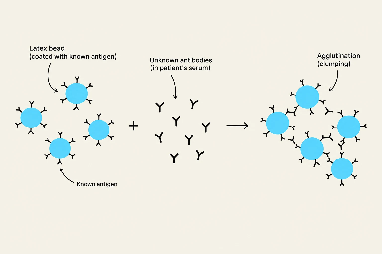
Passive agglutination, also known as indirect or particle agglutination, involves coating a carrier particle (such as latex, gelatin, or synthetic beads) with a known antigen. When the patient's serum (containing unknown antibodies) is added, agglutination occurs only if the specific antibodies are present.
One common clinical application is the Rheumatoid Factor (RF) Test. In this test, latex beads are coated with IgG (the known antigen). If the patient's serum contains Rheumatoid Factor—an autoantibody (specifically anti-IgM) that targets the Fc portion of IgG—the latex beads will clump.
This diagram illustrates the principle of agglutination, a process where antibodies in a patient's serum bind to antigens on latex beads, causing them to clump together. This reaction is used in diagnostic tests to detect the presence of specific antibodies or antigens.
Passive Hemagglutination follows the same principle, but uses Red Blood Cells (RBCs) as the carrier particles instead of latex. An example is the Rubella test, where RBCs are coated with Rubella antigens to detect antibodies in a patient's serum.
Reverse Passive Agglutination
In "reverse" techniques, the roles are swapped: the antibodies are placed on the carrier particles, and the unknown antigen is sought from the patient specimen.
Reverse Passive Agglutination: Uses latex or other particles coated with known antibodies to detect bacterial or fungal antigens, such as Cryptococcus neoformans in cerebrospinal fluid (CSF).
Reverse Passive Hemagglutination: Uses RBCs coated with known antibodies. A clinical example is the detection of Hepatitis B "s" antigen (HBsAg), where anti-HBs antibodies are attached to RBCs to catch the antigen in an unknown sample.
Coagglutination
Coagglutination is a specialized form of reverse agglutination that utilizes bacteria as the inert carrier particle. Staphylococcus aureus is frequently used because its cell wall contains a high concentration of Protein A. Protein A naturally binds to the Fc portion of IgG, leaving the Fab portions of the antibody free to interact with the unknown antigen in the patient specimen.
Inhibition Techniques
Sometimes, the absence of a reaction is actually the indicator of a positive result. This is the basis of inhibition assays.
Agglutination Inhibition
This method relies on competition between a particulate antigen and a soluble antigen for a limited number of antibody binding sites. If the patient's sample contains the target antigen, it will "occupy" the antibodies first, preventing them from binding to the carrier particles. Therefore, a lack of agglutination indicates a positive reaction. A classic example is the β-HCG pregnancy test.
Hemagglutination Inhibition
Similar to the above, this technique uses the inhibition of RBC clumping to detect specific substances, often used in viral or bacterial identification.
Exam Tip: In agglutination inhibition tests, remember that the interpretation is "inverted."
Agglutination present = Negative result.
No agglutination = Positive result.
Challenges in Interpretation: False Results
Laboratory professionals must be wary of several phenomena that can lead to incorrect diagnoses:
Cross-reactivity: When an antibody reacts with an antigen that is structurally similar to the target but is not the intended target.
Rheumatoid Factor: As noted earlier, RF can cause false positive reactions in various agglutination tests.
Prozone Phenomenon: This occurs when there is an excess of antibodies relative to the antigen. The abundance of antibodies prevents the formation of a large, interconnected lattice, resulting in a false negative.
Postzone Phenomenon: This occurs when there is an excess of antigen relative to the antibody.
To avoid these errors, the "Zone of Equivalence" must be reached, where the proportions of antigen and antibody are optimal for forming large, visible complexes.
Precipitation Techniques
While agglutination involves particulate antigens, precipitation involves the combination of soluble antigens with soluble antibodies to produce insoluble complexes.
Light Scattering: Turbidimetry and Nephelometry
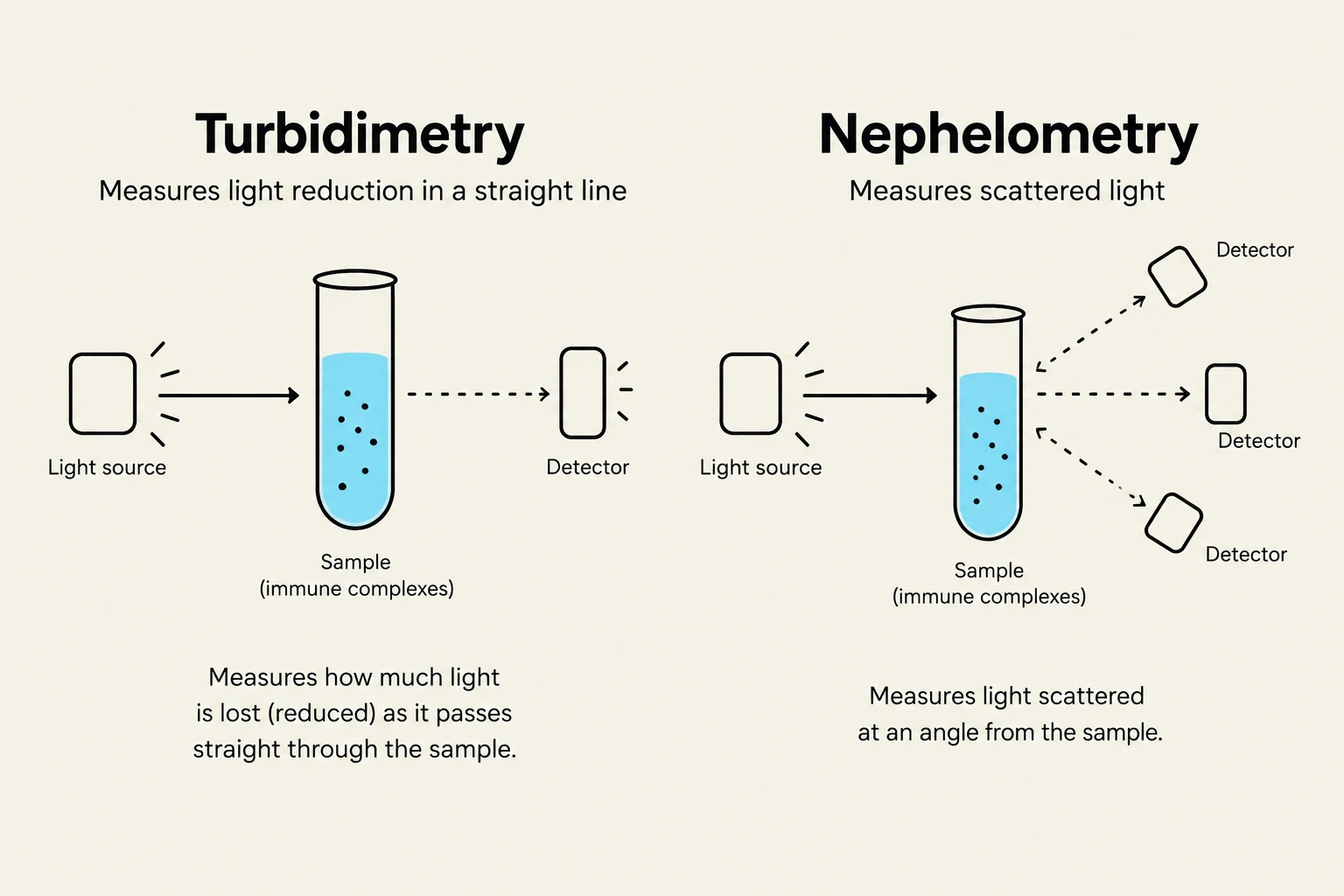
When soluble antigens and antibodies interact in a liquid solution, the solution becomes cloudy (turbid). This cloudiness can be measured using two primary methods:
Turbidimetry: Measures the decrease in light intensity as light passes directly through the solution.
Nephelometry: Measures light scattered at a specific angle from the incident beam. Nephelometry is generally more sensitive than turbidimetry and is used to quantify serum proteins like immunoglobulins and complement components.
This diagram illustrates the principles of turbidimetry and nephelometry, two methods used to measure the concentration of particles in a sample by analyzing how light interacts with it. Turbidimetry measures the reduction in light intensity as it passes straight through the sample, while nephelometry measures the light scattered at an angle.
Passive Immunodiffusion
In these techniques, precipitation occurs within a support medium, typically a gel. The rate of diffusion is influenced by temperature, gel viscosity, and particle size.
Radial Immunodiffusion (Mancini Method): A single diffusion method where reagent antibodies are mixed with an antigen. The antigen diffuses outward, and the resulting ring of precipitation is measured. The diameter of the ring is directly proportional to the antigen concentration.
Ouchterlony Double Diffusion: A method where both the antigen and the antibody diffuse through the gel from separate wells. By observing the patterns where the diffusion fronts meet, clinicians can identify the relationship between antigens:
Identity: An arc of identity forms when the two antigens are the same.
Nonidentity: Crossed lines form when the antigens are completely different.
Partial Identity: A "spur" forms when one antigen shares some, but not all, epitopes with another.
These patterns are vital in clinical settings for fungal identification and diagnosing autoimmune diseases.
Keep reading for free.
Create an account to finish this reading and make it part of your study routine.