Acute Knee and Ankle Injuries: Mechanisms, Diagnosis, and Management
This article explores the biomechanics of acute knee and ankle injuries, focusing on common mechanisms like valgus and rotational forces, hyperextension, and direct blows. It details clinical indicators such as hemarthrosis, physical examination tests like the Lachman and Anterior Drawer tests, and discusses complicati
Acute musculoskeletal injuries are common in both athletic and daily life, often resulting from sudden, high-force events that exceed the physiological limits of connective tissues. Understanding the specific mechanisms of injury—how the force is applied to the joint—is essential for predicting which structures, such as ligaments, tendons, or bones, have been compromised. This article explores the biomechanics of knee and ankle injuries, the clinical indicators of severe damage, and the standard approaches to diagnosis and management.
Mechanisms of Acute Knee Injury
The knee is a complex hinge joint stabilized by several key ligaments. Because of its structure, specific directional forces result in predictable injury patterns.
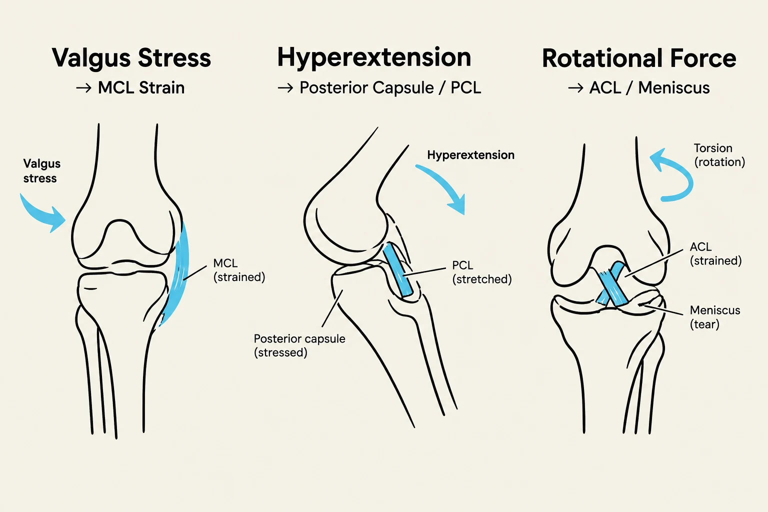
Valgus and Rotational Forces
A valgus stress occurs when a force hits the lateral (outer) side of the knee, pushing the joint inward. This mechanism most commonly results in a Medial Collateral Ligament (MCL) injury, though it can be accompanied by Anterior Cruciate Ligament (ACL) damage.
If the valgus force is combined with external rotation, the risk of a complete ACL rupture increases significantly. Such high-energy mechanisms often involve a "package" of injuries, including the MCL, the lateral meniscus, or even bone bruises on the tibial plateau.
Other Common Mechanisms
Hyperextension: Forcing the knee straight beyond its normal range is a primary mechanism for ACL injuries.
Direct Anterior Tibial Blow: A direct impact to the front of the shin (tibia) can push the bone backward, typically resulting in a Posterior Cruciate Ligament (PCL) tear.
Patellar Dislocation/Subluxation: This occurs when the kneecap moves out of its normal groove, often described by patients as the "knee-cap going out".
Degenerative Changes: In older individuals, minor twisting motions that might not have caused injury in youth may instead lead to degenerative meniscal tears.
Diagrams illustrating three types of knee injuries: valgus stress leading to MCL strain, hyperextension causing posterior capsule and PCL stress, and rotational force resulting in ACL strain and meniscus tear.
Anatomy and Clinical Indicators of ACL Injury
The ACL is a critical intra-articular ligament composed of two distinct functional bundles. The anteromedial bundle is responsible for resisting anterior tibial translation (the shin sliding too far forward), while the posterolateral bundle primarily resists rotation.
Hemarthrosis: A Red Flag
One of the most important clinical indicators of a severe injury is hemarthrosis, which is the rapid bleeding into the joint space. The presence of rapid swelling (within minutes to hours) suggests high-energy damage such as an ACL tear, a peripheral meniscus tear, an osteochondral injury, or a bone fracture. In contrast, injuries like MCL tears or central meniscus tears may cause swelling but are less likely to cause immediate, massive hemarthrosis.
Physical Examination and Diagnosis
To assess for ACL stability, clinicians rely on specific manual tests:
Lachman Test: Performed with the knee at 20–30 degrees of flexion and slightly externally rotated; it is a highly sensitive test for ACL integrity.
Anterior Drawer Test: Performed with the knee at 90 degrees of flexion to check for anterior translation of the tibia.
Exam Tip: When evaluating a patient, always ask: "Did you feel or hear a rip, pop, or tear?" and "Did something come out of place?" These subjective reports are vital for differentiating between a simple sprain and a complete rupture.
Diagrams illustrating the Lachman test and Anterior Drawer test for assessing anterior translation of the tibia.
Complications and Long-term Risks
Acute injuries, if not managed correctly, can lead to permanent structural changes. A classic example is the Unhappy Triad, which involves the simultaneous injury of the ACL, the MCL, and the medial meniscus.
Furthermore, even after successful surgical reconstruction of the ACL, patients face a high risk of long-term issues. There is a significant risk of developing osteoarthritis within 15–20 years following a major knee trauma. For those undergoing ACL reconstruction, the graft is at its weakest between 3 to 6 months post-operation, and returning to high-level sports too early can lead to a four-fold increase in re-injury rates.
Related Injuries: Avulsion Fractures and Ankle Sprains
Avulsion Fractures
An avulsion fracture occurs when a sudden, forceful contraction of a muscle or a violent pull from a ligament/tendon tears a piece of bone away from its attachment site. This results in sudden pain, swelling, and loss of function. Common sites include the ASIS (Anterior Superior Iliac Spine) and the Ischial Tuberosity.
Ankle Sprains and Syndesmosis Injuries
Ankle injuries are among the most common in sports.
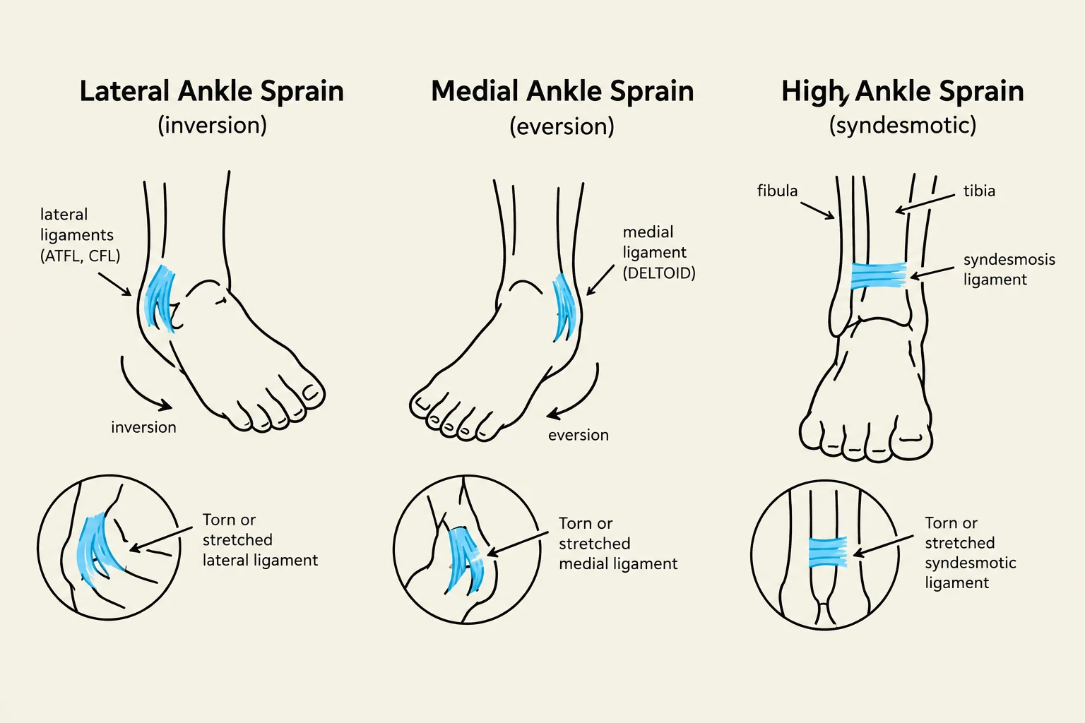
Lateral Ankle Sprains: Caused by sudden inversion or supination, affecting the ATF (Anterior Talofibular), PTF (Posterior Talofibular), and CF (Calcaneofibular) ligaments. These are graded from I (partial rupture) to III (total rupture of two ligaments).
Medial Ankle Sprains: Caused by excessive eversion, affecting the deltoid ligament. These typically take twice as long to heal as lateral sprains.
Syndesmosis Injury (High Ankle Sprain): Resulting from forced external rotation, this involves the ligaments connecting the tibia and fibula.
Osteochondral Injuries (OCI/OCF): These involve damage to the cartilage and underlying bone and often occur alongside ankle sprains, causing stiffness and locking.
Diagram illustrating different types of ankle sprains: lateral (inversion), medial (eversion), and high (syndesmotic), showing the affected ligaments.
Diagnosis and Management Strategies
The standard diagnostic pathway involves a thorough history, physical examination (including palpation), and imaging such as X-rays or MRI.
Treatment Approaches
Treatment is dictated by the severity of the injury:
Conservative Management: For lower-grade sprains or minor injuries, treatment includes load modification, NSAIDs for pain, physiotherapy (focusing on core strength, flexibility, and nerve glides), and proprioception (balance) training.
Bracing and Taping: Often used for Grade I and II ankle sprains to allow for early functional movement.
Surgical Intervention: Necessary for complete syndesmosis ruptures, certain osteochondral fractures (OCF), or complex ACL reconstructions.
Regardless of the method, the ultimate goal is to prevent recurrent instability—such as the feeling of the ankle "giving way"—and to mitigate the long-term risk of osteoarthritis.
Keep reading for free.
Create an account to finish this reading and make it part of your study routine.