Abnormal Electrical Neurotransmission and Epilepsy
This content explains the fundamental mechanisms of epilepsy, focusing on the ionic basis of action potentials, the roles of ion channels and neurotransmitters like GABA and glutamate, and the classification of seizure types. It details how disruptions in neuronal electrical activity lead to seizures and discusses diag
Epilepsy is a complex neurological disorder characterized by a tendency toward recurrent, unprovoked seizures. At its core, epilepsy represents a profound dysfunction of the cerebral cortex, involving transient alterations in brain function that manifest as motor, sensory, autonomic, or psychic symptoms. To understand why these electrical storms occur, we must look closely at the microscopic mechanisms of the neuron: the ion channels that govern the action potential and the neurotransmitters that manage the communication between cells.
The Ionic Basis of the Action Potential
The fundamental unit of neural communication is the action potential. This process is driven by the movement of ions across the neuronal membrane through voltage-gated ion channels (VGIC).
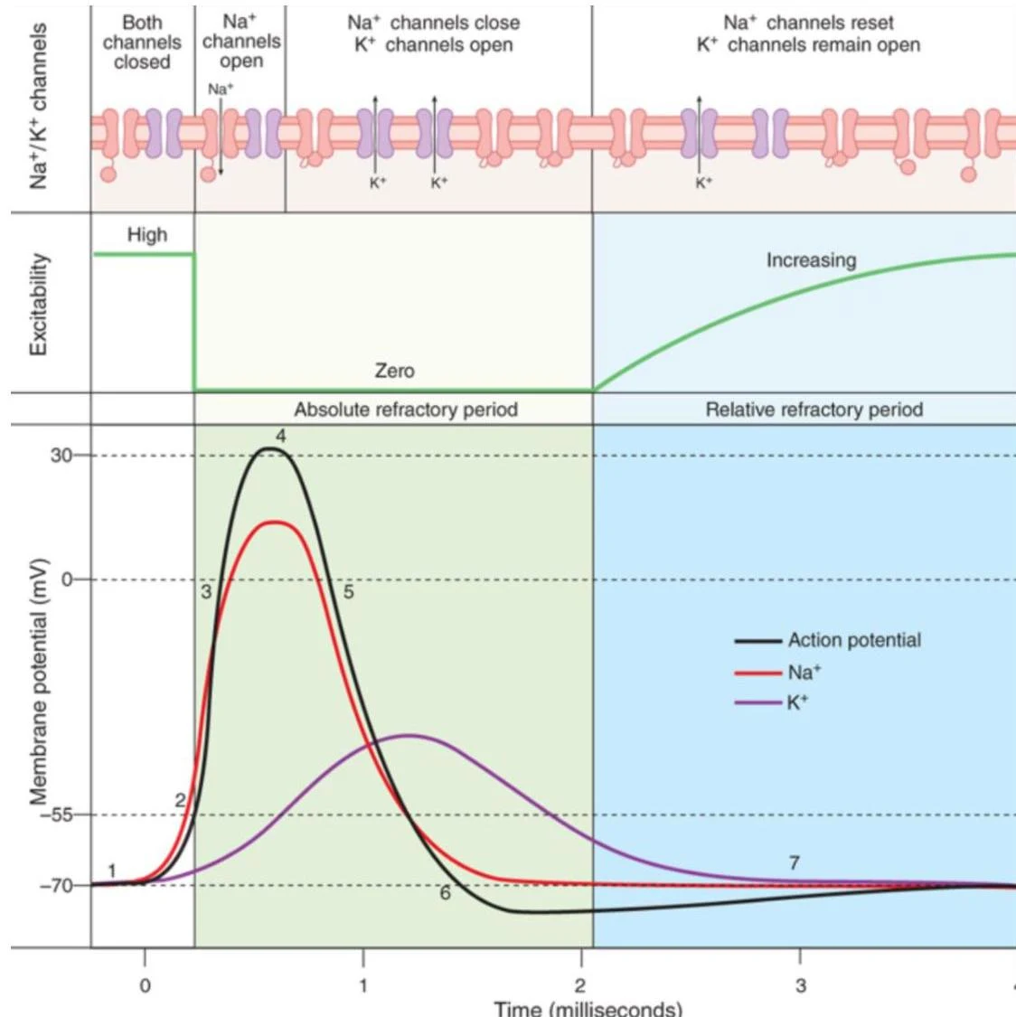
When a neuron receives sufficient stimulation, it undergoes depolarization. This is a rapid process where the membrane potential shifts, triggering the opening of voltage-gated sodium channels (VGSC). As Na+ ions rush into the cell, the membrane potential rises sharply. This is followed by the opening of voltage-gated potassium channels (VGPC), which allows $K^+$ to exit the cell, leading to repolarization or hyperpolarization.
This diagram illustrates the changes in membrane potential during an action potential, showing the roles of Na+ and K+ channels and the refractory periods.
Exam Tip: Remember the sequence of channel activity. VGSC opens faster than VGPC. The opening of VGSC causes depolarization, while the opening of VGPC facilitates repolarization.
Mechanisms of Control: Preventing Repetitive Firing
In a healthy brain, neurons fire in a controlled, isolated manner. Two primary mechanisms prevent neurons from entering the state of uncontrolled, repetitive firing that characterizes a seizure: VGSC inactivation and GABAergic inhibition.
1. VGSC Inactivation and the Refractory Period
Voltage-gated sodium channels do not simply stay open; they transition through three distinct states: resting, open, and inactivated. The transition from the open state to the inactivated state is a critical "braking" mechanism.
The time required for a channel to recover from the inactivated state back to the resting state determines the refractory period. If a neuron attempts to fire too rapidly, the VGSCs will not have sufficient time to recover from inactivation. This imposes a physical "time-limit" on how frequently a neuron can fire. If a group of neurons manages to override these limits, they can engage in repetitive, synchronous firing, which is a hallmark of seizure activity.
2. GABAergic Interneurons and Surround Inhibition
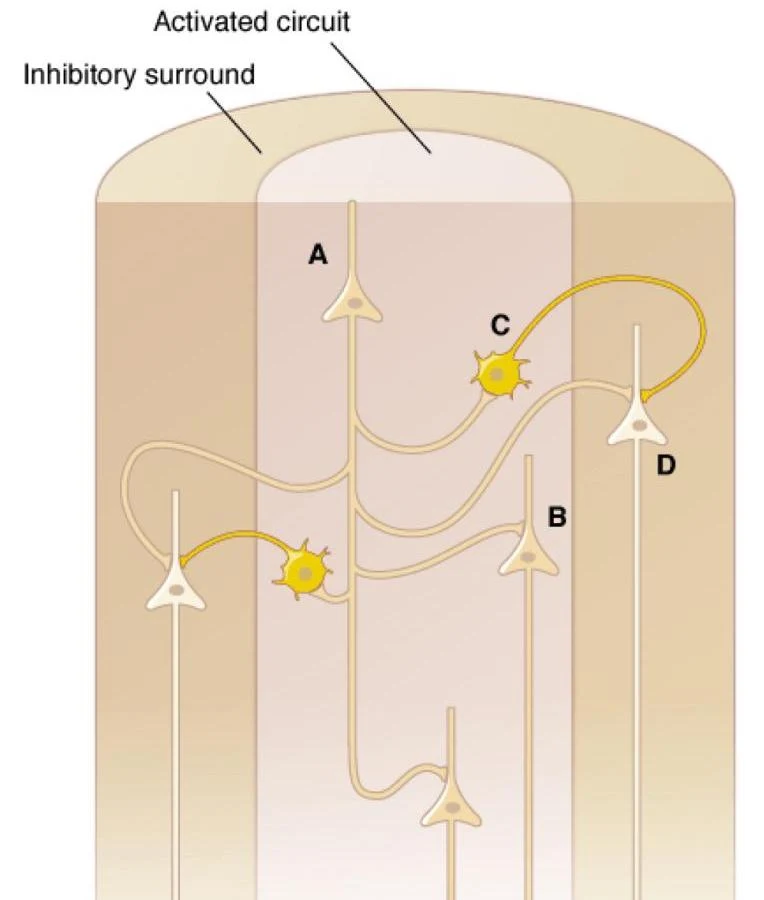
While VGSCs control signal propagation at the single-cell level, GABAergic interneurons control the network level. These inhibitory neurons release GABA (gamma-aminobutyric acid), which acts on GABA type A receptors to suppress electrical activity in surrounding neurons.
This process, known as "surround inhibition," prevents the spread of electrical signals to unnecessary areas and suppresses synchronous discharges. By maintaining this balance, the brain prevents a localized signal from becoming a widespread, synchronized seizure.
Diagram illustrating neural circuits with an activated circuit and an inhibitory surround, showing connections between neurons labeled A, B, C, and D.
Neurotransmitter Imbalance: Glutamate vs. GABA
The balance of the brain relies on the interplay between excitatory and inhibitory neurotransmission.
Excitatory Neurotransmission: Primarily mediated by glutamate. Glutamate acts on ionotropic receptors, specifically AMPA receptors (which mediate fast transmission) and NMDA receptors (which are crucial for calcium influx and synaptic plasticity).
Inhibitory Neurotransmission: Primarily mediated by GABA acting on GABA type A receptors.
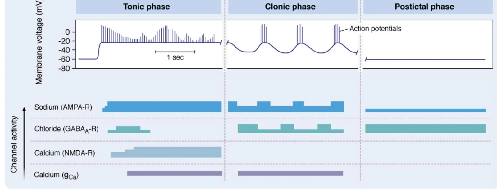
During a tonic-clonic seizure, we see a massive shift in this balance. The tonic phase is characterized by intense sodium influx (via AMPA receptors), while the clonic phase and subsequent postictal depression involve complex shifts in calcium (via NMDA receptors) and chloride (via GABA receptors).
This diagram illustrates the changes in membrane voltage and ion channel activity during the tonic, clonic, and postictal phases of a seizure.
Classification of Seizures
Clinically, seizures are classified based on where they begin in the brain and whether they affect consciousness.
Focal Onset Seizures
These begin in a localized area or group of cells within one cerebral hemisphere.
Focal Aware Seizures: The patient remains conscious (formerly "simple partial").
Focal Impaired Awareness Seizures: The patient's consciousness or contact with reality is affected (formerly "complex partial"). These may include automatisms, such as lip-smacking or repetitive hand movements.
Generalized Onset Seizures
These involve both cerebral hemispheres from the start.
Tonic-Clonic Seizures: These involve a sudden loss of consciousness. They consist of a tonic phase (sustained muscle contraction and limb extension) followed by a clonic phase (rhythmic limb jerking).
Absence Seizures: These are non-motor seizures characterized by brief "staring spells" or loss of consciousness. They are often associated with a specific EEG pattern: generalized 3-Hz spike-and-wave activity, which is driven by abnormal activity in the thalamus via T-type Ca2+ channels.
Unknown Onset
If the beginning of the seizure is not witnessed or cannot be determined, it is classified as an unknown onset seizure.
Clinical Note: A seizure is a single episode, whereas epilepsy is the clinical diagnosis of a disorder characterized by recurrent, unprovoked seizures.
Diagnostic Tools
To differentiate these seizure types, clinicians rely on:
Electroencephalogram (EEG): This records electrical activity from the scalp. It can identify interictal spike discharges (spontaneous spikes between seizures) or Paroxysmal Depolarization Shifts (PDS), which are high-voltage, long-duration depolarizations caused by Na+ and Ca2+ influx.
Brain Imaging (CT, MRI): Used to identify structural causes such as tumors, trauma, or cerebrovascular issues.
Keep reading for free.
Create an account to finish this reading and make it part of your study routine.